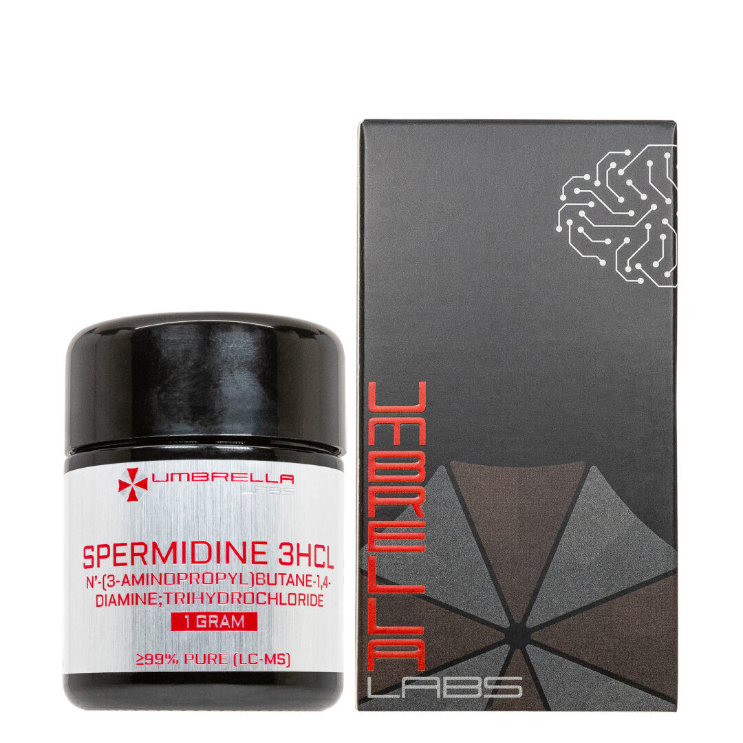
SPERMIDINE 3HCL 30ML LIQUID (20MG/ML, 600MG BOTTLE)
$25.99
Spermidine 3HCL is sold for laboratory research use only. Terms of sale apply. Not for human consumption, nor medical, veterinary, or household uses. Please familiarize yourself with our Terms & Conditions prior to ordering.


Also Available In:
 Capsule Option >> Powder Option >>
- Description
- Additional information
Description
Spermidine 3HCl Nootropic Liquid


| CAS Number | 334-50-9 |
| Other Names | 334-50-9, Spermidine Hydrochloride, N1-(3-Aminopropyl)butane-1,4-diamine trihydrochloride |
| IUPAC Name | N’-(3-aminopropyl)butane-1,4-diamine;trihydrochloride |
| Molecular Formula | C₇H₂₂Cl₃N₃ |
| Molecular Weight | 254.63 |
| Purity | ≥99% Pure (LC-MS) |
| Liquid Availability | 30mL liquid (20mg/mL, 600mg bottle) |
| Powder Availability | 60 capsules (10mg/capsule, 600mg total)
|
| Storage | Store in cool dry environment, away from direct sunlight. |
| Terms | All products are for laboratory developmental research USE ONLY. Products are not for human consumption. |
What is Spermidine 3HCl?
Spermidine is a naturally occurring polyamine compound that has been studied for its potential role in supporting cognitive function, cellular health, and healthy aging. Found throughout the body and in a variety of foods, spermidine is involved in important biological processes such as cell growth, mitochondrial regulation, and the stimulation of autophagy, a process that helps clear damaged cellular materials and maintain proper function. By promoting cellular renewal and reducing the effects of oxidative stress and inflammation, spermidine may contribute to improved neuronal protection and overall brain health. Emerging research suggests that spermidine may help preserve cognitive abilities by supporting pathways associated with memory, learning, and long-term neurological function.
Main Research Findings
1) In a model of diet-induced obese mice, spermidine was found to improve the function of gut microbiota and improve the integrity of the gut barrier.
2) Treatment with spermidine was found to reduce brain aging through the induction of autophagy in a model of SAMP8 mice.
Selected Data
1) The study conducted by the research team of Ma et al investigated the effects of spermidine supplementation on obesity-associated metabolic dysfunction, intestinal barrier impairment, inflammation, and gut microbiota alterations. The researchers designed a comprehensive experimental approach involving human dietary data analysis, multiple mouse models, microbiome sequencing, biochemical measurements, molecular analyses, and cellular experiments to determine the mechanisms by which spermidine influences metabolic health. Since spermidine is a naturally occurring polyamine obtained from dietary sources and produced by intestinal microorganisms, the authors examined whether changes in spermidine availability could influence obesity development and related complications [1].
The first component of the study involved an analysis of human clinical and dietary data obtained from the National Health and Nutrition Examination Survey (NHANES) database. Information from 5,799 individuals was collected, and spermidine intake was estimated using dietary information combined with a previously established polyamine content database. The researchers focused specifically on 1,908 participants without diabetes to examine associations between daily spermidine intake and metabolic indicators such as body mass index (BMI), waist circumference, fasting glucose, fasting insulin, hemoglobin A1c (HbA1c), and the homeostatic model assessment of insulin resistance (HOMA-IR). Statistical correlations were performed using Pearson correlation analysis to determine whether natural dietary spermidine intake was related to obesity-associated metabolic parameters [1].
To investigate the effects of spermidine experimentally, the researchers used eight-week-old male C57BL/6J mice. The mice were separated into dietary groups and maintained for sixteen weeks under different feeding conditions. One group received a normal chow diet, while another group received a high-fat diet designed to induce obesity and metabolic dysfunction. Additional high-fat diet groups were supplemented with different doses of spermidine through drinking water, allowing the researchers to determine whether spermidine administration could reverse or prevent diet-induced obesity effects. Body weight, food intake, and water intake were regularly monitored throughout the experiment. After treatment, metabolic tissues including liver, adipose tissue, intestine, and fecal samples were collected for further analysis.
Several metabolic assessments were performed to evaluate the impact of spermidine on obesity-related complications. Body composition was examined by measuring fat accumulation, and tissue samples from the liver and epididymal white adipose tissue were evaluated using histological staining techniques. Hematoxylin and eosin (H&E) staining was used to examine tissue structure, liver lipid accumulation, and adipocyte size. Glucose metabolism was measured through glucose tolerance tests and insulin tolerance tests, while circulating glucose, insulin levels, and HOMA-IR calculations were used to determine insulin sensitivity. Inflammation within metabolic tissues was examined by measuring macrophage accumulation using F4/80 staining and evaluating inflammatory signaling pathways, including NF-κB activation.
Because the researchers hypothesized that spermidine affected obesity through improvements in intestinal health, extensive analyses of gut barrier function were performed. Colon length was measured as an indicator of intestinal health, and intestinal permeability was assessed using fluorescein isothiocyanate (FITC)-dextran testing, which measures the movement of molecules across the intestinal barrier. Plasma lipopolysaccharide (LPS) levels were also measured because increased circulating LPS indicates bacterial product leakage from the gut and contributes to systemic inflammation. Additional intestinal assessments included staining methods to measure mucus-producing goblet cells and mucin secretion, which are important components of the protective intestinal barrier [1].
To determine the molecular pathways affected by spermidine, the researchers performed transcriptomic and protein analyses on intestinal tissues. RNA sequencing was used to identify changes in gene expression caused by high-fat feeding and spermidine treatment. The analysis focused on pathways associated with epithelial function, inflammation, lipid metabolism, and autophagy. Quantitative PCR was used to confirm changes in genes related to autophagy, tight junction proteins, mucin production, and inflammatory responses. Western blot analyses measured proteins involved in intestinal barrier function, including Claudin-1 and Occludin, as well as autophagy-related proteins such as LC3B and Beclin-1. Apoptosis-related proteins, including Bax, Bcl2, and Caspase-3, were also examined to determine whether spermidine protected intestinal cells from damage.
Additional experiments were performed using Caco-2 intestinal epithelial cells to investigate cellular mechanisms. These cells were treated with spermidine to determine its direct effects on intestinal barrier proteins, autophagy activity, and inflammatory signaling. Researchers also examined whether spermidine acted through Toll-like receptor 4 (TLR4), a receptor involved in detecting bacterial LPS and activating inflammation. Experiments using TLR4 knockout mice and pharmacological TLR4 inhibition helped determine whether suppression of this pathway contributed to spermidine’s protective effects.
The study also explored the relationship between spermidine and the gut microbiome. Cecal microbiota samples were analyzed using high-throughput 16S rRNA sequencing to determine bacterial composition and diversity. Bioinformatic analysis identified changes in bacterial populations caused by obesity and spermidine supplementation. To determine whether gut bacteria were required for spermidine’s effects, researchers used antibiotic treatment to deplete intestinal microbiota. They also performed fecal microbiota transplantation (FMT), transferring microbiota from spermidine-treated mice into obese mice to evaluate whether altered bacterial communities alone could improve metabolic outcomes. Finally, short-chain fatty acid (SCFA) concentrations were measured because these bacterial metabolites are important regulators of intestinal barrier function and metabolism. Together, these methods allowed the researchers to examine spermidine’s effects at the organism, tissue, cellular, and microbial levels [1].
2) The study performed by researchers Xu et al was designed to investigate the potential anti-aging and neuroprotective effects of the naturally occurring polyamines spermidine and spermine, with a specific focus on their influence on cognitive function, autophagy activation, mitochondrial regulation, oxidative stress, inflammation, and neuronal survival. Since aging-related cognitive decline is associated with decreased autophagy, impaired mitochondrial function, oxidative damage, and neurodegeneration, the researchers used a well-established accelerated aging mouse model to examine whether polyamine supplementation could counteract these biological changes. The study compared the effects of spermidine and spermine with rapamycin, a known inducer of autophagy, to determine whether similar cellular pathways contributed to their protective effects [2].
The animal model used in this investigation consisted of three-month-old male senescence-accelerated mouse prone 8 (SAMP8) mice and senescence-accelerated mouse resistant 1 (SAMR1) mice. SAMP8 mice are commonly used as a model of accelerated aging because they naturally develop age-associated characteristics, including learning deficits, memory impairment, oxidative stress, and neurodegenerative changes. SAMR1 mice served as the normal aging control group because they do not develop the same rapid aging phenotype. All animals were housed under specific pathogen-free laboratory conditions and maintained according to established animal care guidelines. The experimental groups included SAMR1 control mice, untreated SAMP8 mice, and SAMP8 mice receiving spermidine, spermine, or rapamycin treatments. Spermidine and spermine were administered orally through drinking water at a concentration of 2 mmol/L, while rapamycin was administered intragastrically at a dose of 0.78 mg/kg/day. Treatments were continued for eight weeks to evaluate the long-term effects of polyamine supplementation on brain aging and neurological function [2].
Following treatment, behavioral experiments were performed to determine whether spermidine and spermine improved age-associated cognitive impairment. The first behavioral assessment was the novel object recognition test (ORT), which evaluated recognition memory based on the natural tendency of mice to explore unfamiliar objects. During the initial phase, mice were placed in a testing chamber without objects to allow them to adapt to the environment. On the following day, two identical objects were placed in the chamber, and the amount of time each mouse spent exploring them was recorded. During the final test phase, one familiar object was replaced with a new object, and memory ability was assessed by measuring preference for the novel object. Researchers calculated the discrimination index by comparing exploration time of the novel object with total exploration time. Improved recognition of the novel object indicated stronger memory retention.
The researchers also conducted an open field test (OFT) to measure locomotor activity, exploratory behavior, and anxiety-related responses. Mice were individually placed in a square testing arena divided into multiple sections and allowed to move freely for five minutes while their behavior was recorded by an overhead camera. Researchers measured movement patterns, distance traveled within the central squares, time spent in the inner area, and exploratory activities such as standing and grooming behaviors. Because aging-related neurological decline can reduce exploratory behavior and increase anxiety-like responses, improvements in these measurements were interpreted as indicators of enhanced neurological function. The arena was cleaned between trials using ethanol to prevent odor-based behavioral influences.
After behavioral testing, brain tissues were collected for molecular and biochemical analyses. Oxidative stress was evaluated by measuring markers associated with cellular damage and antioxidant defense. Malondialdehyde (MDA), an indicator of lipid oxidation caused by reactive oxygen species, was measured to determine the degree of oxidative injury. Superoxide dismutase (SOD) activity was also examined because SOD acts as an antioxidant enzyme that protects cells from oxidative damage. ATP concentrations were measured to evaluate mitochondrial energy production because mitochondrial dysfunction is a major characteristic of aging neurons. These biochemical measurements helped determine whether polyamine treatment improved cellular metabolism and protected the aging brain from oxidative stress [2].
The study also used Western blot analysis to investigate proteins involved in autophagy, mitochondrial function, inflammation, apoptosis, and neuronal health. Brain tissue samples were processed to extract proteins, which were separated using SDS-polyacrylamide gel electrophoresis and transferred onto membranes for antibody detection. Autophagy activity was assessed by measuring phosphorylated AMP-activated protein kinase (p-AMPK), Beclin-1, LC3, and p62. Increased p-AMPK, Beclin-1, and LC3 combined with reduced p62 levels were interpreted as evidence of enhanced autophagic activity. Since mitochondrial balance is essential for healthy aging, proteins involved in mitochondrial fusion and function were analyzed, including mitofusin-1 (MFN1), mitofusin-2 (MFN2), dynamin-related protein 1 (DRP1), and cytochrome c oxidase IV (COX IV) [2].
To determine whether spermidine and spermine protected neurons from degeneration, the researchers examined apoptosis and inflammatory pathways. Western blotting measured apoptosis-related proteins, including the protective protein Bcl-2 and the cell death-associated proteins Bax and Caspase-3. TUNEL staining was performed on brain sections to directly detect apoptotic cells in areas such as the hippocampus and cortex. Inflammation was analyzed by measuring proteins associated with inflammatory signaling, including NLRP3, interleukin-1β (IL-1β), and interleukin-18 (IL-18).
Finally, the researchers examined neuroprotective and synaptic markers, including nerve growth factor (NGF), brain-derived neurotrophic factor (BDNF), postsynaptic density protein 93 (PSD93), and postsynaptic density protein 95 (PSD95), to determine whether polyamine treatment supported neuronal communication and plasticity. Overall, these methods allowed the researchers to comprehensively evaluate whether spermidine and spermine could reduce brain aging through improvements in autophagy, mitochondrial health, oxidative balance, and neuronal survival [2].
Discussion
1) The results of the study by researchers Ma et al demonstrated that spermidine supplementation provided significant protection against obesity-related metabolic dysfunction by improving intestinal barrier integrity, reducing inflammation, activating protective cellular pathways, and altering gut microbiota composition. The researchers first examined whether spermidine intake was associated with obesity-related characteristics in humans by analyzing data from the NHANES. They found that daily dietary spermidine intake was negatively correlated with several obesity and metabolic disease indicators, including BMI, waist circumference, fasting glucose, fasting insulin, HbA1c, and insulin resistance measured by the HOMA-IR index [1].
Individuals with higher markers of obesity and impaired glucose regulation generally consumed lower amounts of spermidine. Similar patterns were observed in mice, where high-fat diet-induced obese animals displayed significantly reduced fecal spermidine levels compared with lean animals. Fecal spermidine concentrations in mice were negatively associated with body weight, fat mass, blood glucose, insulin levels, and insulin resistance, suggesting that reduced spermidine availability may be linked to the development of obesity-related metabolic abnormalities.
After establishing this association, the researchers investigated whether spermidine supplementation could improve metabolic outcomes in high-fat diet-induced obese mice. Mice receiving spermidine through their drinking water for sixteen weeks experienced a significant dose-dependent reduction in body weight gain compared with untreated obese mice. Importantly, this decrease occurred without major differences in food or water consumption, indicating that the protective effect was not caused by reduced caloric intake. Analysis of body composition revealed that the reduction in weight was primarily due to decreased fat accumulation rather than loss of lean tissue. Spermidine-treated mice showed reductions in epididymal white adipose tissue mass and smaller adipocyte size. Although liver weight was not significantly changed, histological examination revealed that spermidine reduced excessive lipid accumulation in the liver, suggesting protection against obesity-associated fatty liver development. Spermidine treatment also improved glucose metabolism, as treated mice demonstrated improved glucose tolerance, increased insulin sensitivity, reduced circulating insulin levels, and lower HOMA-IR scores compared with untreated obese mice. These metabolic improvements were associated with enhanced insulin signaling in metabolic tissues, including the liver and adipose tissue [1].
The researchers also found that spermidine reduced obesity-associated inflammation. High-fat diet feeding increased macrophage accumulation in both liver and adipose tissue, which was indicated by elevated F4/80 staining and the formation of inflammatory crown-like structures in fat tissue. Spermidine administration significantly reduced macrophage infiltration and decreased activation of inflammatory pathways, including NF-κB signaling. These findings suggested that spermidine reduced chronic inflammation, a major contributor to insulin resistance and metabolic disease. Additional results showed that spermidine increased thermogenic activity in brown adipose tissue by enhancing markers associated with energy expenditure, such as uncoupling protein 1 (UCP1). This indicated that spermidine may contribute to improved metabolic regulation by increasing energy utilization in addition to reducing inflammation and fat accumulation.
A major finding of the study was that spermidine significantly improved intestinal barrier function, which is commonly disrupted during obesity. High-fat diet-fed mice displayed shorter colon lengths, increased intestinal permeability, and elevated circulating lipopolysaccharide (LPS) levels, indicating damage to the intestinal barrier and increased movement of bacterial products into circulation. Spermidine treatment reversed these effects by increasing colon length, lowering intestinal permeability, and decreasing plasma LPS concentrations. Histological examination showed that spermidine increased mucus-producing goblet cells and enhanced mucin secretion, strengthening the protective barrier of the intestine. RNA sequencing revealed that spermidine reversed many high-fat diet-induced changes in gene expression, particularly genes involved in epithelial development, cell signaling, inflammation, and intestinal function. Spermidine increased expression of genes related to tight junction formation, including Claudin and Tjp family genes, while decreasing inflammatory markers [1].
Further investigation revealed that spermidine’s ability to improve gut barrier integrity was associated with activation of autophagy, a cellular recycling and repair mechanism. High-fat diet feeding reduced intestinal autophagy markers, while spermidine increased expression of autophagy-associated genes and proteins such as LC3B and Beclin-1. Spermidine also increased tight junction proteins, including Claudin-1 and Occludin, while reducing proteins involved in cell death pathways. Similar effects were observed in Caco-2 intestinal epithelial cells, confirming that spermidine directly enhanced intestinal cell protection. In experiments involving LPS-induced intestinal injury, spermidine prevented colon damage, decreased inflammatory markers, reduced intestinal permeability, and restored barrier-related proteins. These results indicated that spermidine preserved intestinal health by promoting autophagy, reducing apoptosis, and maintaining epithelial integrity [1].
The researchers next examined whether spermidine influenced the gut microbiome. High-fat diet feeding disrupted microbial composition, increasing bacteria associated with metabolic dysfunction while decreasing beneficial bacterial populations. Spermidine supplementation shifted the gut microbiota profile toward one resembling healthier lean mice. Specifically, spermidine increased beneficial SCFA-producing bacteria, particularly the Lachnospiraceae NK4A136 group. This bacterial group showed strong associations with improved intestinal barrier function, including lower intestinal permeability, reduced LPS levels, increased colon length, and higher tight junction gene expression. Spermidine also increased levels of beneficial microbial metabolites, especially acetate, propionate, and butyrate, which are known to support gut barrier function and reduce inflammation.
To determine whether the microbiota changes were necessary for spermidine’s benefits, researchers depleted gut bacteria using antibiotics. After microbiota removal, spermidine was no longer able to improve intestinal barrier function, glucose tolerance, insulin resistance, or metabolic outcomes, demonstrating that gut bacteria were required for many of its protective effects. Conversely, fecal microbiota transplantation from spermidine-treated mice into obese mice partially transferred these benefits. Recipient mice showed improved intestinal integrity, decreased LPS levels, enhanced insulin signaling, and better metabolic profiles, even without direct spermidine supplementation. These findings confirmed that spermidine-induced changes in gut bacteria played an important role in mediating its anti-obesity effects [1].
Finally, the researchers investigated the role of TLR4 signaling, a pathway activated by bacterial LPS that contributes to inflammation and metabolic dysfunction. Spermidine reduced activation of TLR4 and its downstream signaling molecule MyD88 in both intestinal tissues and cultured intestinal cells. Experiments using TLR4-deficient mice showed that loss of TLR4 produced similar improvements to spermidine treatment, including improved gut barrier function and insulin sensitivity. However, spermidine provided little additional benefit when TLR4 signaling was already absent, suggesting that inhibition of this pathway was a major mechanism underlying spermidine’s effects. Overall, the study concluded that spermidine protects against diet-induced obesity by strengthening the intestinal barrier, promoting autophagy, reducing inflammatory TLR4 signaling, and restoring beneficial gut microbiota populations. These results suggest that spermidine supplementation may represent a potential strategy for improving obesity-related metabolic disorders [1].
2) The results of the study by Xu et al demonstrated that long-term administration of the natural polyamines spermidine and spermine significantly reduced multiple characteristics of brain aging by improving cognitive function, enhancing autophagy, restoring mitochondrial balance, decreasing oxidative damage, preventing neuronal apoptosis, reducing inflammation, and supporting synaptic function. Using the SAMP8 model, which naturally develops aging-related neurological impairments, the researchers compared untreated aging mice with mice receiving spermidine, spermine, or the autophagy-inducing compound rapamycin. The findings showed that spermidine and spermine produced protective effects similar to rapamycin, suggesting that their anti-aging benefits were strongly associated with the activation of autophagy and the maintenance of neuronal health [2].
The first major finding of the study was that spermidine and spermine improved memory retention and cognitive performance in aging mice. Cognitive function was assessed using the novel ORT, which measures recognition memory by determining whether mice can distinguish between familiar and unfamiliar objects. Untreated SAMP8 mice displayed significant memory impairment compared with the normal-aging SAMR1 control mice, demonstrated by reduced exploration of the novel object and a lower discrimination index.
After treatment with spermidine, spermine, or rapamycin, SAMP8 mice spent more time investigating the new object, showing that memory recognition ability was restored. These results suggested that polyamine treatment was able to reverse or reduce cognitive decline associated with accelerated aging. In the OFT, which measured locomotor activity and anxiety-related behavior, untreated SAMP8 mice displayed reduced exploratory behavior and less movement within the central area of the testing environment. Mice treated with spermidine and spermine showed increased activity, greater distance traveled in the inner squares, and increased time spent exploring the central region. These behavioral improvements indicated that polyamine supplementation enhanced neurological function and reduced aging-associated behavioral deficits [2].
The researchers next examined whether spermidine and spermine influenced oxidative stress, a major contributor to brain aging and neurodegeneration. Aging is associated with the accumulation of reactive oxygen species that damage cellular components, including proteins, lipids, and DNA. The untreated SAMP8 mice showed increased levels of MDA, a marker of lipid peroxidation and oxidative injury, along with decreased activity of the antioxidant enzyme SOD. Treatment with spermidine, spermine, and rapamycin significantly decreased MDA levels while increasing SOD activity within brain tissue. These findings demonstrated that polyamines strengthened antioxidant defenses and protected neurons from oxidative damage. By restoring the balance between reactive oxygen species production and antioxidant activity, spermidine and spermine helped create a healthier cellular environment that could support neuronal survival during aging [2].
The study also found that spermidine and spermine improved neuronal communication by increasing the expression of proteins involved in synaptic function and neuroplasticity. Aging and neurodegenerative conditions are commonly associated with synaptic loss, which contributes to impaired learning and memory. The researchers measured important neuronal markers, including NGF, BDNF, PSD93, and PSD95. Untreated SAMP8 mice exhibited reduced expression of these proteins, suggesting impaired neuronal support and weakened synaptic connections. Following treatment with spermidine, spermine, or rapamycin, expression levels of NGF, BDNF, PSD93, and PSD95 were significantly increased. These changes indicated that polyamine supplementation supported synaptic stability, promoted neuronal survival, and enhanced pathways involved in learning and memory formation. The improvement in these molecular markers was consistent with the behavioral improvements observed during memory testing.
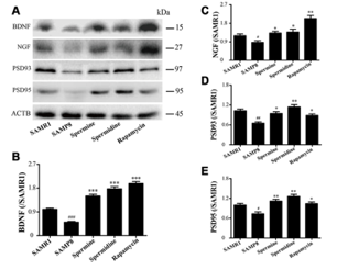
Figure 1: A) Western blot of NGF, PSD93, PSD95, and BDNF; B) changes in BDNF levels; C) changes in NFG; D) changes in PSD93; and E) changes in PSD95.
Another important result was that spermidine and spermine restored mitochondrial function in aging brains. Mitochondrial dysfunction is considered one of the major hallmarks of aging because mitochondria provide the energy required for neuronal activity and regulate processes such as oxidative stress and cell survival. Untreated SAMP8 mice showed abnormal expression of mitochondrial regulatory proteins and reduced ATP production, indicating impaired mitochondrial performance. Treatment with spermidine and spermine increased the levels of mitochondrial fusion proteins MFN1 and MFN2, which are responsible for maintaining mitochondrial structure and function. The treatments also influenced DRP1, a protein involved in mitochondrial fission, and increased COX IV, an important marker of mitochondrial respiration. Additionally, ATP concentrations increased in treated mice, suggesting improved energy production. These findings demonstrated that polyamines helped preserve mitochondrial balance and provided neurons with the energy necessary for normal function [2].
The researchers further investigated whether the protective effects of spermidine and spermine were connected to autophagy, a cellular recycling process responsible for removing damaged proteins and organelles. The results showed that untreated SAMP8 mice had impaired autophagic activity, demonstrated by decreased levels of autophagy-related proteins. Spermidine and spermine treatment increased phosphorylation of AMPK, a major regulator that promotes autophagy. In addition, treatment increased the expression of Beclin-1 and LC3-II, proteins involved in autophagosome formation and autophagic activity, while decreasing levels of p62, a protein that accumulates when autophagy is impaired. These changes indicated that spermidine and spermine restored normal autophagic function in aging brain tissue. Since rapamycin produced similar results, the findings supported the idea that polyamine-induced autophagy activation was a key mechanism responsible for delaying brain aging.
The study also revealed that spermidine and spermine protected neurons by reducing apoptosis, or programmed cell death. Aging SAMP8 mice displayed increased expression of pro-apoptotic proteins, including Bax and cleaved Caspase-3, along with reduced levels of the protective anti-apoptotic protein Bcl-2. TUNEL staining confirmed that untreated aging mice had increased numbers of apoptotic cells in important brain regions such as the hippocampus and cortex. Treatment with spermidine, spermine, and rapamycin reversed these changes by decreasing Bax and Caspase-3 expression while increasing Bcl-2 levels. The number of TUNEL-positive cells was also greatly reduced, indicating improved neuronal survival. These results suggested that polyamines prevented excessive neuron loss during aging by regulating pathways responsible for cell death [2].
Finally, spermidine and spermine significantly decreased brain inflammation associated with aging. Untreated SAMP8 mice showed increased activation of inflammatory pathways, including elevated levels of NLRP3 inflammasome proteins, IL-1β, and IL-18. These inflammatory factors are known to contribute to neuronal damage and cognitive decline. After treatment with spermidine, spermine, or rapamycin, the expression of these inflammatory markers was sharply reduced, demonstrating that polyamines suppressed neuroinflammation. Overall, the results showed that spermidine and spermine delayed brain aging through multiple interconnected mechanisms: activating AMPK-mediated autophagy, restoring mitochondrial function, improving energy production, decreasing oxidative stress, preventing neuronal apoptosis, reducing inflammation, and preserving synaptic proteins involved in memory. The researchers concluded that these natural polyamines may represent promising compounds for protecting against age-related cognitive decline and neurodegenerative changes [2].
Disclaimer
**LAB USE ONLY**
*This information is for educational purposes only and does not constitute medical advice. THE PRODUCTS DESCRIBED HEREIN ARE FOR RESEARCH USE ONLY. All clinical research must be conducted with oversight from the appropriate Institutional Review Board (IRB). All preclinical research must be conducted with oversight from the appropriate Institutional Animal Care and Use Committee (IACUC) following the guidelines of the Animal Welfare Act (AWA).
Citations
[1] Ma L, Ni Y, Wang Z, et al. Spermidine improves gut barrier integrity and gut microbiota function in diet-induced obese mice. Gut Microbes. 2020;12(1):1-19. doi:10.1080/19490976.2020.1832857
[2] Xu TT, Li H, Dai Z, et al. Spermidine and spermine delay brain aging by inducing autophagy in SAMP8 mice. Aging (Albany NY). 2020;12(7):6401-6414. doi:10.18632/aging.103035
Characteristics of Spermidine 3HCl
Spermidine 3HCl is a nootropic compound originally discovered by the Dutch scientist, Anton Van Leeuwenhoek. Spermidine is found in the sperm and is derived from two polyamines, putrescine and spermine. Polyamines are considered unique due to their ability to attach to a multitude of different molecules. That being said, polyamines are shown to be involved in the regulation of DNA stability and cellular functions such as apoptosis, proliferation, and growth. This involvement indicates that polyamines function in a similar fashion as growth factors in cellular division. Since it is composed of two different polyamines, spermidine HCL can be considered essential to healthy growth and development. Additional benefits of spermidine 3HCl, include increased longevity, anti-aging, and improved health of the reproductive system.
Since spermidine plays an integral role in cell metabolism and functioning, the compound is typically found in all eukaryotic cells. Spermidine also acts as an essential growth factor when found in various types of bacteria. The compound is readily available in many whole foods such as a multitude of vegetables, soybean products, wheat germ cereals, and aged cheeses, however, the spermidine levels in these foods are low. Animal-based studies have found that food-extracted supplementation has shown to be more efficient in eliciting benefits in subjects.
Longevity Benefits of Spermidine 3HCl
While typical supplements work by filling in gaps in nutrients, spermidine 3HCl works through its active ingredients that manipulate the molecular pathways involved in aging. One of the primary examples of this is spermidine’s ability to induce autophagy. Autophagy naturally occurs in cells in order to regulate cellular metabolism. Furthermore, it is considered to be the process of cellular rejuvenation through the involvement of the TOR kinase pathway. Autophagy is also responsible for the balance of nutrients during times of increased cellular stress such as fasting. This explains why compounds such as spermidine that mimic fasting and calorie restriction induce autophagy. Overall, this mechanism is thought to promote longevity and anti-aging as autophagy effectively limits inflammation and oxidative stress.
Research related to spermidine’s ability to combat inflammation and oxidative stress has found that supplementation with the compound is able to protect DNA from oxidation and mitochondrial DNA from age-related mutations. That being said, aging, degeneration, and certain diseases, such as cancer, are correlated to the length of telomeres. Telomeres act as end caps to chromosomes and while they naturally shorten with age, the rate of shortening directly impacts longevity. Both telomere length and instability of the mitochondrial genome are used to measure age in changing individuals, however, animal-based research has found that administration of spermidine actively inhibits more than 50% of typical aging hallmarks (https://longevity.technology/news/spermidine-a-geroprotector-supplement/).
Current research has found that in addition to improving overall health and longevity, spermidine is able to prevent neurodegeneration and many age-related diseases such as heart disease or cancer. The compound’s neuroprotective effects that work to treat inflammation, ischemia, and oxidative stress are also related to spermidine’s potential ability to improve memory and cognition. Through autophagy, spermidine and dissolving amyloid-𝜷 proteins in the brain. This is important to note considering that amyloid-𝜷 proteins tend to build into plaques that cause dementia and Alzheimer’s Disease. Early studies have supported this claim and reported that spermidine is able to prevent memory loss in animals with induced dementia.
Fertility Benefits of Spermidine 3HCl
Another interesting benefit of Spermidine 3HCl is its potential to improve fertility in both male and female, aged animals. Female animals are able to have children until they reach menopause. Research has found that the onset of menopause can be accelerated through the accumulation of reactive oxygen species (ROS) in the ovaries. ROS is a form of oxidative stress that has been shown to damage oocytes and cause cellular degeneration in the reproductive system. ROS can also limit fertility by causing abnormal development of the female germline stem cells (FGSCs). Researchers Li et. Al suggested that abnormalities in FGSCs caused by oxidative stress could be prevented by administering spermidine. Results of their research found the FGSCs treated with spermidine experienced increased autophagy and stem cell production (https://pubmed.ncbi.nlm.nih.gov/31319247/).
Studies have found that spermidine levels are lower in infertile male subjects. While male animals do not experience reproductive aging as rapidly as female animals do, spermidine is still capable of improving male fertility as well. Sperm production is highly influenced by different hormones, and is notably decreased by the presence of cortisol. Researchers Bendera et. Al found that administration of spermidine for 30 days could drastically lower cortisol levels in subjects. Results also found that the compound was able to reduce levels of oestradiol, a hormone typically linked to decreased sexual functioning in males. The researchers also observed increased testosterone and DHEAs, allowing them to conclude that spermicide is effective in terms of male reproductive health and functioning (https://longevity.technology/lifestyle/everything-you-need-to-know-about-spermidine/).
The nootropics sold by Umbrella Labs are sold for laboratory research only. The description above is not medical advice and is for informative purposes only.
Spermidine 3HCL is sold for laboratory research use only. Terms of sale apply. Not for human consumption, nor medical, veterinary, or household uses. Please familiarize yourself with our Terms & Conditions prior to ordering.


| File Name | View/Download |
| 2026-03-24-Umbrella-Labs-Spermidine-Certificate-of-Analysis-COA.pdf |  |
VIEW CERTIFICATES OF ANALYSIS (COA)
Additional information
| Weight | 1 oz |
|---|---|
| Dimensions | 1.25 × 1.25 × 4 in |
| Options |