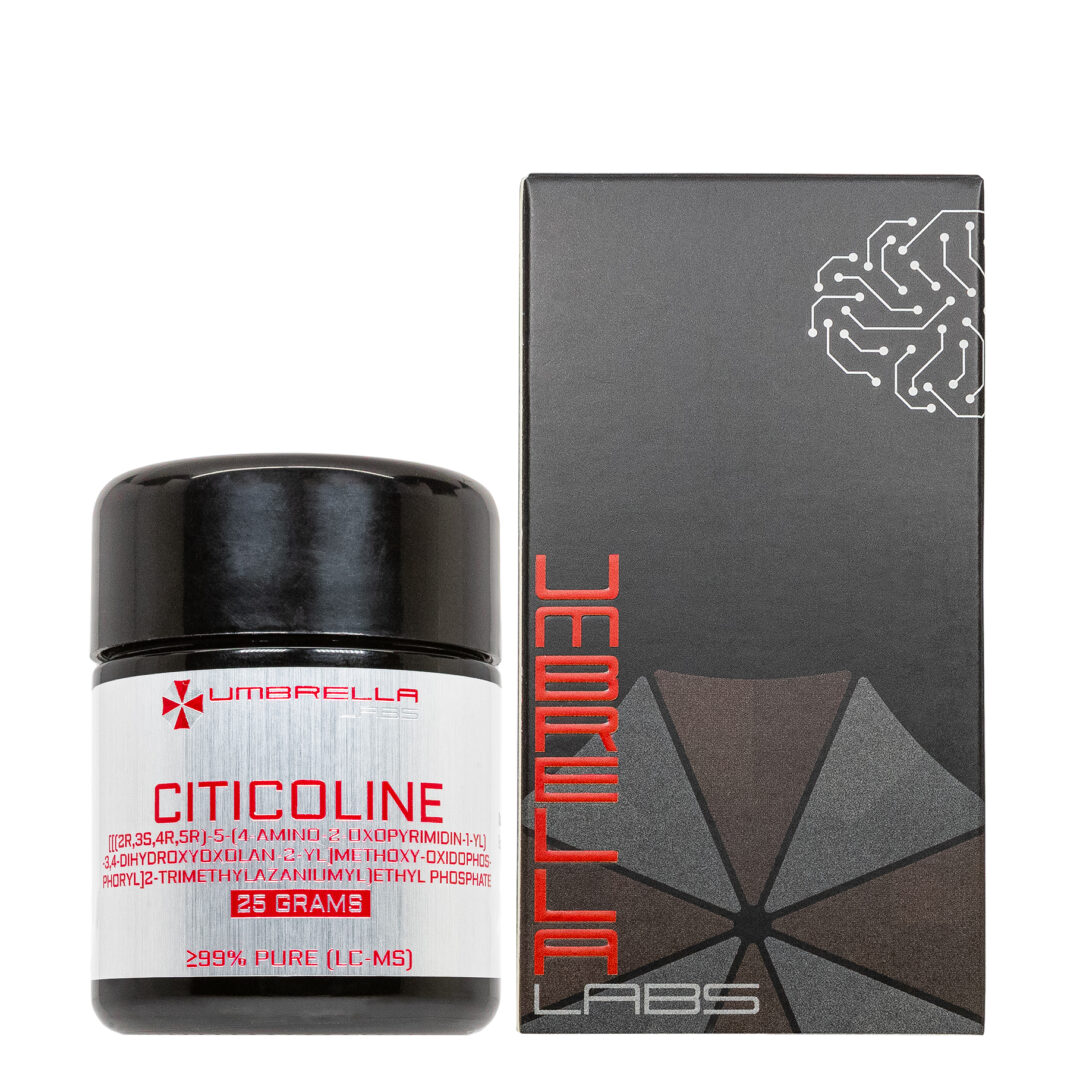
COMPOUND 7P POWDER (1 GRAM)
$101.98
Compound 7P is sold for laboratory research use only. Terms of sale apply. Not for human consumption, nor medical, veterinary, or household uses. Please familiarize yourself with our Terms & Conditions prior to ordering.

Also Available In:
 Liquid Option >>
- Description
- Additional information
Description
Compound 7P Nootropic Powder


| CAS Number | 1890208-58-8 |
| Other Names | Acetamide, 2-[(2-methoxyphenyl)[(4-methylphenyl)sulfonyl]amino]-N-(4-methoxy-3-pyridinyl)- |
| IUPAC | 2-(2-methoxy-N-(4-methylphenyl)sulfonylanilino)-N-(4-methoxypyridin-3-yl)acetamide |
| Molecular Formula | C₂₂H₂₃N₃O₅S |
| Molecular Weight | 441.5 |
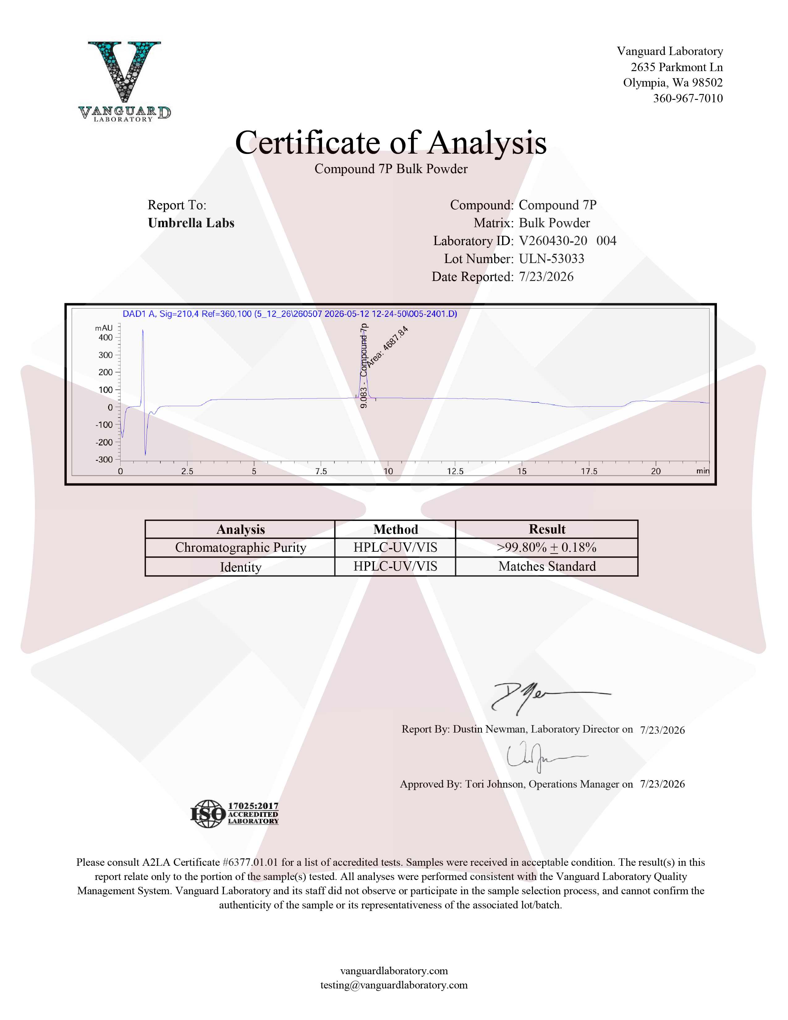
| Purity | ≥99% Pure (LC-MS) |
| Liquid Availability | 30mL liquid (50mg/mL, 1500mg bottle) |
| Powder Availability | 1 gram |
| Storage | Store in a dry, cool, dark place. For best preservation, store at 4°C or colder away from bright light. |
| Terms | All products are for laboratory developmental research USE ONLY. Products are not for human consumption. |
What is Compound 7P?
Compound 7P is a small-molecule nootropic compound that has attracted scientific interest for its potential neuroprotective and neuroregenerative properties. Compound 7P has been studied largely in the context of neuronal growth, axonal regeneration, and repair following nervous-system injury. Prior research has identified Compound 7P as a candidate capable of promoting neurite outgrowth, a process through which developing or injured neurons establish and restore connections with other cells. Studies have also noted the ability of the compound to inhibit cancer-related gene expression. The activity of this compound has generated interest in its potential applications for neurological conditions involving neuronal damage, degeneration, or impaired axonal connectivity.
Main Research Findings
1) In an animal model of Alzheimer’s disease, Compound 7p was found to reduce cognitive and memory deficits.
2) In an animal model of optic nerve injury, Compound 7p was shown to promote neuronal outgrowth and regeneration.
Selected Data
1) The study by Yu et al investigated a series of pyranone-carbamate compounds designed to simultaneously inhibit butyrylcholinesterase (BuChE) and reduce neuroinflammatory activity. The researchers synthesized 19 novel derivatives, designated 7a–7s, using a 3-hydroxylpyranone scaffold connected to a carbamate-containing aromatic system through a styrene-based linker. The design was based on the premise that BuChE inhibition could increase cholinergic signaling while suppression of neuroinflammation could address another important component of Alzheimer’s disease pathology. The authors had previously identified 3-hydroxylpyranone as a useful anti-Alzheimer’s scaffold with anti-inflammatory, antioxidant, neuroprotective, and brain-access properties. They incorporated a carbamate group because this chemical functionality can interact with cholinesterases, while the styrene unit was selected as a linker between the pyranone and carbamate portions. The resulting compounds were evaluated through a progression of chemical, biochemical, cellular, computational, pharmacokinetic, toxicological, and behavioral experiments, ultimately identifying Compound 7p as the lead molecule [1].
After synthesis, the chemical structures of the target compounds were characterized using ^1H and ^13C nuclear magnetic resonance spectroscopy and high-resolution mass spectrometry. Melting-point analysis and high-performance liquid chromatography were used to assess compound purity, and all compounds used for biological testing had purities greater than 95%. Compound 7p received additional structural confirmation through X-ray crystallographic analysis, which verified the trans configuration of the vinyl bond produced during the Heck reaction. The experimental chemistry was monitored using thin-layer chromatography, while NMR, HRMS, melting-point, and HPLC measurements were performed using the instrumentation described by the investigators. These analytical procedures ensured that biological testing was performed with structurally verified and sufficiently pure compounds.
The first biological evaluation examined inhibition of acetylcholinesterase and BuChE. The researchers tested the compounds against electric-eel AChE (eeAChE), equine-serum BuChE (eqBuChE), and human-serum BuChE (huBuChE) using a modified Ellman assay. Tacrine served as the reference inhibitor. Initial testing was performed at 20 μM for AChE and 10 μM for BuChE, and compounds producing more than 50% inhibition were subsequently subjected to IC₅₀ determination. The assay used a 100 μL reaction mixture containing the test compound, DTNB, enzyme, and phosphate-buffered saline at pH 8.0. After incubation at 37°C, acetylthiocholine or butyrylthiocholine was added as the enzymatic substrate. Absorbance was measured at 412 nm using a microplate reader, and IC₅₀ values were calculated. For the more detailed kinetic evaluation of 7p, different concentrations of the compound and butyrylthiocholine were examined, with absorbance monitored every minute for 10 minutes. Dixon and Cornish-Bowden plots were subsequently constructed by linear regression to establish the inhibition mechanism and calculate inhibition constants [1].
Anti-neuroinflammatory activity was evaluated using BV-2 mouse microglial cells and an LPS-induced nitric oxide production model. Before measuring inflammation, an MTT cell-viability assay was performed to determine concentrations that did not substantially compromise cell viability and thereby prevent false-positive results. BV-2 cells were seeded at 1 × 10^4 cells per well and exposed to the compounds for 24 hours before MTT was added. Formazan was dissolved with DMSO and absorbance was measured at 490 nm. Because no significant viability differences were observed at 10 μM for the compounds or hydrocortisone, the investigators selected 10 μM for the subsequent neuroinflammation experiments. For nitric oxide measurement, BV-2 cells were seeded at 5 × 10^4 cells per well and incubated for 24 hours. Test compounds were then added for one hour before LPS was introduced at a final concentration of 1 μg/mL. After another 24 hours, culture supernatants were collected and nitric oxide levels were determined using a commercial nitric oxide assay based on the Griess methodology. Hydrocortisone served as the reference anti-inflammatory compound [1].
The investigators next evaluated whether 7p possessed properties appropriate for a central nervous system drug. SwissADME was used to predict physicochemical properties, drug-likeness, and blood-brain barrier permeability for compounds 7a–7s. Compound 7p was then experimentally assessed using a parallel artificial membrane permeation assay designed to model BBB penetration. The test compound and reference drugs were dissolved in DMSO, diluted in PBS, and placed in a donor plate containing a porcine-brain-lipid membrane. The donor and acceptor plates were assembled and incubated at 25°C for 10 hours. The concentration transferred to the acceptor compartment was measured spectrophotometrically, and the apparent permeability coefficient was calculated. Six commercially available drugs with established BBB characteristics were used to validate the assay.
Safety and behavioral efficacy were subsequently examined in mice. For acute toxicity, 16 ICR mice, equally divided by sex, were randomly assigned to a control group receiving 0.5% CMC-Na or a Compound 7p group receiving a single oral dose of 1000 mg/kg. Animals were observed for 14 days for changes in behavior, food and water consumption, body weight, and survival. At the end of the observation period, animals were sacrificed and the heart, liver, spleen, and brain were examined histologically after hematoxylin-eosin staining [1].
For cognitive testing, C57BL/6J mice were assigned to control, scopolamine, rivastigmine, and two Compound 7p groups, with eight animals per group. Scopolamine was administered intraperitoneally at 5 mg/kg from days 1–26 to produce cognitive impairment. Beginning on day 11, rivastigmine at 1 mg/kg or Compound 7p at 1 or 5 mg/kg was administered orally once daily. Behavioral testing occurred on days 21–26 using the Morris water maze. Animals underwent five days of training to locate a hidden platform, followed by a probe trial in which the platform was removed and the number of platform crossings was recorded. Animal trajectories were recorded by camera and analyzed [1].
2) The research team of Ku et al. investigated whether small-molecule compounds could stimulate neuronal outgrowth and promote axonal regeneration using a phenotypic, cell-based screening approach. The initial screen was designed to identify compounds capable of activating pathways involved in neurite extension. Compounds were tested at 10 μM in P19 embryonic carcinoma cells that had been transiently transfected with a NeuroD2 expression construct. NeuroD2 induced neuronal differentiation in the P19 cells, resulting in the formation of neurite-like projections that could be measured. Compounds that produced more than a twofold increase in neurite outgrowth compared with the DMSO control were considered active. This primary screening process generated 185 confirmed hits, representing a 0.11% hit rate [2].
The compounds identified during the initial screen were then evaluated in primary rat hippocampal neurons to determine whether their effects could be reproduced in a more physiologically relevant neuronal model. Primary hippocampal and cortical neurons were isolated from embryonic day 18 Sprague–Dawley rats. The hippocampus and cerebral cortex were separately removed, mechanically dissociated, washed, filtered, and plated for cell culture. After 24 hours, the neurons were treated with the test compounds for an additional 48 hours. Neuronal morphology was subsequently examined through indirect immunofluorescence staining using βIII-tubulin as a neuronal marker. A high-content imaging system captured fluorescence from four fields per well, while specialized neuronal profiling software was used to quantify neurite development. This secondary screen identified several compounds containing a common sulfonamidoacetamide scaffold. Compound 1 was selected for additional development because it produced pronounced neurite elongation in hippocampal neurons and showed similar activity in cortical neurons. However, its poor water solubility and limited metabolic stability prevented direct in vivo testing. The researchers therefore used structure–activity relationship optimization, with neurite outgrowth in primary cortical neurons serving as the primary biological endpoint [2].
After identifying Compound 7p as a promising optimized analogue, its effects were investigated in retinal neurons. Retinal ganglion cells were isolated from postnatal day 7 Sprague–Dawley rats using a commercial isolation kit. The cells were seeded onto coverslips coated with poly-D-lysine and laminin at a density of 200,000 cells per well. Compound 7p was administered at concentrations of 0, 2, 10, and 20 μM. Following 72 hours of treatment, the cells were fixed and stained with the SMI-312 anti-neurofilament antibody to visualize axonal structures. Neuronal growth was assessed according to axonal length, with cells categorized into groups measuring less than 50 μm, 50–100 μm, 100–200 μm, or greater than 200 μm. Retinal ganglion cells were identified based on their morphology and cell-body size, with cells having a soma diameter greater than 10 μm included in the analysis. Cell viability was additionally evaluated by counting cells across nine consecutive, nonoverlapping microscopic fields.
The researchers ultimately assessed Compound 7p in vivo using an optic nerve crush model to determine whether its effects on neuronal growth could translate into axonal regeneration. Male Sprague–Dawley rats approximately eight weeks old and weighing 200–250 g were maintained under controlled environmental conditions with regulated temperature, humidity, and a 12-hour light/dark cycle. Animals were randomly assigned to either a sham-operated control group or an optic nerve crush group receiving Compound 7p. Under anesthesia, the retrobulbar optic nerve was exposed and mechanically crushed with forceps for 15 seconds at approximately 2 mm behind the optic head. Animals in the experimental group subsequently received intravitreal injections of 0.22 μg of Compound 7p in 5 μL per eye every four days for three weeks, while sham-operated animals received saline. At the end of treatment, the eyes and optic nerves were collected, fixed in paraformaldehyde, cryoprotected, embedded, sectioned, and immunostained. GAP-43 was used as a marker of actively growing axons, and ImageJ software was used to quantify GAP-43-positive axons at distances of 0.5, 1.0, and 1.5 mm from the optic nerve crush site [2].
Discussion
1) The study performed by Yu et al successfully generated a library of 19 pyranone-carbamate derivatives and identified Compound 7p as the most promising molecule based on its combination of BuChE inhibition and anti-neuroinflammatory activity. Chemical characterization confirmed the expected structures of the synthesized derivatives, and all compounds subjected to biological testing had purities above 95%. X-ray crystallography provided additional confirmation for Compound 7p and demonstrated the trans configuration of its vinyl linkage. The synthetic strategy therefore successfully produced structurally diverse compounds that allowed the researchers to systematically examine how modifications to the carbamate substituent, pyranone moiety, and linker affected biological activity [1].
The researchers then modified the pyranone portion and linker to optimize the activity of the piperidine-containing scaffold. Removal of the pyranone 6-methyl group produced 7l, which inhibited eqBuChE with an IC₅₀ of 8.10 nM and huBuChE with an IC₅₀ of 6.93 nM. Replacing the methyl group with hydroxymethyl generated 7m, which produced eqBuChE and huBuChE IC₅₀ values of 5.22 and 16.53 nM, respectively. Altering the 3-position substituent also affected activity. Larger substituents, such as those incorporated into 7n and 7o, reduced BuChE potency, whereas replacing the larger O-benzyl group with a smaller O-methyl group generated Compound 7p. This modification substantially improved activity, giving 7p an eqBuChE IC₅₀ of 4.68 ± 1.39 nM and a huBuChE IC₅₀ of 9.12 ± 0.57 nM. By comparison, 7g produced values of 15.29 and 38.68 nM, respectively. These findings established 7p as the strongest BuChE inhibitor within the series.
The anti-neuroinflammatory experiments showed that the compounds also influenced LPS-induced nitric oxide production in BV-2 microglial cells. The LPS-treated group showed a substantial increase in NO production relative to untreated controls, confirming successful induction of the inflammatory model. The compounds subsequently reduced NO production to varying degrees. Compound 7k demonstrated the strongest NO inhibition, reducing NO production by 35.41% at 10 μM. However, 7k possessed relatively weak BuChE activity. Compound 7p was considered more therapeutically attractive because it produced a more balanced dual profile. At 10 μM, 7p reduced NO production by 28.82%, and its activity was comparable to hydrocortisone. The SAR analysis indicated that converting the 6-methyl group to hydroxymethyl, changing the 3-O-benzyl substituent to O-methyl, and hydrogenating the vinyl linker could improve anti-inflammatory activity. Compound 7p therefore represented the compound with the most favorable balance between BuChE inhibition and suppression of inflammatory NO production [1].
Mechanistic studies provided additional evidence explaining the strong BuChE activity of 7p. Enzyme kinetic analysis using Dixon and Cornish-Bowden plots demonstrated that Compound 7p behaved as a mixed-type BuChE inhibitor. The calculated inhibition constants were 3.15 nM for binding to the free enzyme and 3.84 nM for binding to the enzyme-substrate complex. These results suggested that 7p could interact with both the peripheral anionic site and catalytic active site of BuChE. Molecular docking supported this interpretation. The carbamate portion of 7p was positioned near the catalytic active site and formed hydrogen-bond interactions involving Ser-198 and His-438. Additional hydrogen bonds involved the pyranone carbonyl and oxygen atoms interacting with Asp-70 and Thr-120. In contrast, docking to AChE indicated that the narrower active-site groove prevented 7p from fully accessing the catalytic site, leaving it primarily associated with the peripheral anionic site. This structural difference provided a proposed explanation for the compound’s strong selectivity toward BuChE [1].
Computational ADME analysis indicated that most compounds possessed favorable drug-like properties according to Lipinski’s rule of five. Compound 7p was subsequently tested experimentally for BBB permeability using PAMPA-BBB. The assay was successfully validated using six reference drugs, producing a strong correlation between experimental and reported permeability values. Compound 7p demonstrated a permeability coefficient of 5.70 × 10⁻⁶ cm/s, placing it within the study’s high-BBB-permeability category. This result was important because a compound intended to treat Alzheimer’s disease must be capable of reaching the central nervous system. The acute toxicity study also produced favorable findings. ICR mice receiving 1000 mg/kg of 7p showed no deaths or abnormal behavioral effects during the 14-day observation period. Body weights generally increased over the study, and histological examination revealed no obvious pathological abnormalities in the heart, liver, spleen, or brain. The authors therefore reported an acute oral LD₅₀ greater than 1000 mg/kg under the tested conditions.
Finally, Compound 7p demonstrated cognitive benefits in the scopolamine-induced mouse model. The scopolamine group developed clear learning and memory deficits, with an escape latency of 52.63 seconds compared with 20.82 seconds in controls and only 1.5 platform crossings compared with 3.4 in controls. Treatment with rivastigmine reduced escape latency to 31.15 seconds. Compound 7p produced comparable improvements, reducing latency to 30.24 seconds at 1 mg/kg and 29.12 seconds at 5 mg/kg. Platform crossings also increased with 7p treatment, reaching 3.3 crossings at 1 mg/kg and 3.5 crossings at 5 mg/kg, compared with 3.0 crossings for rivastigmine. The mice maintained an overall upward body-weight trajectory throughout the behavioral experiment, supporting the absence of obvious adverse effects during treatment. The results therefore demonstrated that 7p could counteract scopolamine-induced deficits in spatial learning and memory [1].
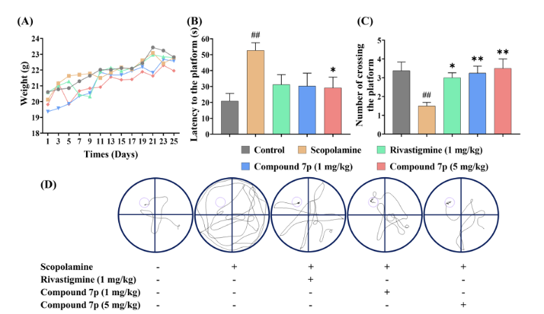
Figure 1: Changes in parameters measured during the Morris Water Maze test across the different experimental groups.
Overall, the study identified Compound 7p as a multifunctional lead molecule combining potent and selective BuChE inhibition, anti-neuroinflammatory activity, predicted drug-likeness, high BBB permeability, favorable acute tolerability, and behavioral efficacy in a mouse model of cognitive impairment. The authors consequently proposed 7p as a promising candidate for further investigation as a potential Alzheimer’s disease therapy, however further studies are warranted [1].
2) The results of the study by Ku et al. showed that their phenotypic screening and subsequent structure–activity optimization successfully identified Compound 7p as a promising molecule for stimulating neurite extension and axonal regeneration. The initial screening campaign evaluated approximately 170,000 synthetic compounds for their ability to enhance neurite outgrowth in NeuroD2-transfected P19 cells. Compounds were tested at 10 μM, and molecules that produced more than a twofold increase in neurite growth relative to DMSO-treated controls were classified as hits. A total of 185 compounds were confirmed as active, corresponding to a 0.11% hit rate. Secondary evaluation in primary rat hippocampal neurons further narrowed the candidates and identified a chemically related group containing a sulfonamidoacetamide scaffold. Compound 1 demonstrated the strongest activity within this group. At concentrations of 10–20 μM, it produced approximately a threefold increase in neurite outgrowth, whereas its activity was diminished at concentrations below 1 μM. Similar neurite-elongating effects were also observed in primary cortical neurons, supporting the sulfonamidoacetamide structure as a useful foundation for further optimization [2].
Although Compound 1 showed substantial biological activity, its unfavorable physicochemical characteristics limited its potential for in vivo application. The researchers therefore performed structure–activity relationship studies to develop analogues with improved biological and pharmacological properties. Among the optimized compounds, Compound 7p emerged as the most effective pyridine-containing analogue. In addition to retaining strong neurite outgrowth activity, 7p demonstrated substantially improved physicochemical and metabolic characteristics compared with Compound 1. Its calculated log P was 2.43, considerably lower than the 3.76 value reported for Compound 1, indicating reduced lipophilicity. Compound 7p also demonstrated a major improvement in metabolic stability. Following incubation with an S9 metabolic fraction, 61.2% of 7p remained compared with only 0.7% of Compound 1. The combination of maintained biological activity, reduced lipophilicity, and improved metabolic stability led the investigators to select 7p for subsequent biological experiments [2].
The researchers next examined whether the neurite-promoting effects of 7p extended to retinal neurons. Retinal neuronal cells were treated with Compound 7p at concentrations between 2 and 20 μM for 72 hours before axonal structures were visualized using anti-neurofilament immunostaining. Axons were classified according to their length to quantify changes in neuronal growth. Compound 7p enhanced axonal extension across the different length categories, with the strongest response observed at 10 μM. Importantly, treatment did not significantly reduce the number of viable retinal neuronal cells. Across the 2–20 μM concentration range, there was no significant difference in total live-cell numbers compared with controls. Therefore, the increased axonal growth appeared to reflect enhanced neuronal extension rather than selective survival of a small subset of cells. These findings provided additional evidence that 7p could promote axonal development in multiple neuronal systems without producing an obvious cytotoxic effect under the tested conditions.
The strongest evidence for axon-regenerative activity came from the in vivo optic nerve crush experiment. Rats with experimentally injured optic nerves received intravitreal injections of Compound 7p at 0.22 μg per eye every four days for three weeks. After treatment, optic nerve tissue was analyzed using GAP-43 immunostaining, with GAP-43 serving as a marker of actively growing axons. Animals treated with 7p showed greater extension of GAP-43-positive axons beyond the injury site compared with PBS-treated injured controls. Increased numbers of GAP-43-positive axons were detected at 500, 1,000, and 1,500 μm from the crush center. The difference reached statistical significance at 1,500 μm, where the injured control group demonstrated approximately 0.22 ± 0.081 GAP-43-positive axons compared with 0.70 ± 0.20 in the Compound 7p-treated group. These results suggested that the neurite-promoting effects observed in cultured neuronal systems could translate into enhanced axonal regeneration following optic nerve injury in vivo [2].
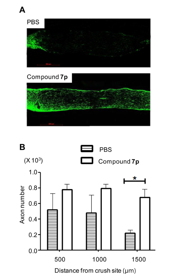
Figure 2: Changes in axonal regeneration in the optic nerve crush site following treatment with Compound 7p.
Overall, the study identified Compound 7p as the most promising derivative generated through the screening and optimization process. Its effects were demonstrated across several neuronal models, including primary hippocampal, cortical, and retinal neurons, while its substantially greater metabolic stability distinguished it from the original Compound 1. Treatment with 7p also increased GAP-43-positive axon extension following optic nerve injury, providing experimental evidence of enhanced axonal regeneration in an injured central nervous system pathway. However, the precise molecular mechanism underlying this effect was not established. The study also did not demonstrate improvements in functional neurological outcomes, cognition, or other behavioral measures. Therefore, the findings support Compound 7p as an experimental neuroregenerative and axon-promoting compound, but they do not establish it as a clinically validated nootropic or therapeutic agent [2].
Disclaimer
**LAB USE ONLY**
*This information is for educational purposes only and does not constitute medical advice. THE PRODUCTS DESCRIBED HEREIN ARE FOR RESEARCH USE ONLY. All clinical research must be conducted with oversight from the appropriate Institutional Review Board (IRB). All preclinical research must be conducted with oversight from the appropriate Institutional Animal Care and Use Committee (IACUC) following the guidelines of the Animal Welfare Act (AWA).
Citations
[1] Yu C, Liu X, Ma B, et al. Novel anti-neuroinflammatory pyranone-carbamate derivatives as selective butyrylcholinesterase inhibitors for treating Alzheimer’s disease. J Enzyme Inhib Med Chem. 2024;39(1):2313682. doi:10.1080/14756366.2024.2313682
[2] Ku JM, Park K, Lee JH, et al. Discovery, Optimization, and Biological Evaluation of Sulfonamidoacetamides as an Inducer of Axon Regeneration. J Med Chem. 2016;59(10):4676-4687. doi:10.1021/acs.jmedchem.6b00015
Compound 7P and Axonal Regeneration
Compound 7P is a nootropic compound originally developed to assist in neurite outgrowth and cognitive enhancement. Compound 7P is currently being researched to determine the role it could potentially play in terms of axon regeneration. Degeneration of axons can occur for a number of reasons, most common being trauma or injury, neurological diseases, and aging. Unfortunately, once the neurons are damaged they cannot repair themselves or regenerate without additional assistance.
Some of the most common methods of growth promotion focus on understanding the specific mechanisms of action and the manipulation of various signaling pathways. The most prevalent mechanisms typically involve epigenetic regulation of gene transcription, development-dependent transcription, and the mTOR and STAT3 signaling pathways. Researchers Park et. Al have discovered that altering the expression of these pathways can effectively lead to axon regeneration in the CNS.
Building off the work of Park et. Al, Ku et. Al have attempted to find additional modalities that will result in axon growth. The research team was able to visually assess and measure all in vitro neurite outgrowth through the use of phenotypic cell-based screens. It’s important to note that using these phenotypic small screen methods of measurement allow researchers to identify different types of protein targets and explain the mechanisms of various signaling pathways. Furthermore, being able to track axon elongation via phenotypic small screen provides an opportunity to more accurately pharmacologically manipulate the different factors assisting axon regeneration.
Use of Compound 7P to Promote Optic Nerve Regeneration
The research team of Ku et. Al was able to use phenotypic screening in order to identify compound 7P. They concluded that compound 7P was able to improve neurite outgrowth in the primary neurons of the hippocampus, cerebral cortex, and retina. Further testing found that in animal models, compound 7P was able to induce regeneration of the optic nerves in cases of injury.
The study initially began with treating retina neuronal cells with doses of either 2 or 20 μM of compound 7P. The cells were then incubated for 72 hours and treated with paraformaldehyde and an anti-neurofilament antibody in order to identify and measure axonal changes. During observation of the cultures the researchers were able to measure cell body size and morphology in order to identify retinal ganglion cells (RGCs). That being said, results of this initial study found that compound 7P led to significant axon growth. Axonal growth increased the most when the cells were treated with a 10 μM dose of the compound.
The second portion of the study was to determine if treatment with compound 7P would elicit the same effect in the RGCs of injured optic nerves as it did in healthy RGCs. The crushed optic nerves were treated with either a placebo, or a daily dose of 0.22 μg per eye, for 3 weeks. Following treatment, the animals were euthanized and cross sections of the optic nerves were stained with an anti-GAP43 antibody so researchers could efficiently identify and measure axonal growth. Results found that when the crushed optic nerves were treated with compound 7P the axons grew remarkably farther past the lesion epicenter than the nerves treated with a placebo. The results of this study suggest that further research should be conducted in order to determine how compound 7P can potentially increase axon growth and treat neuronal damage throughout the central nervous system (https://pubs.acs.org/doi/10.1021/acs.jmedchem.6b00015#).
Effects of Compound 7P Against Cervical Cancer
In addition to its ability to promote axonal regeneration, researchers Wang et. Al examined the potential of compound 7P to combat cervical cancer. The researchers originally hypothesized that electron-withdrawing substituents, such as compound 7P, have more potent anti-cancer qualities than electron-donating groups. Results of the study were based on the histological assessment of a mouse xenograft model inoculated with HeLa cells. It was reported that compound 7P exhibited the highest anti-cancer activity by actively inhibiting the growth of cervical tumors. Excision of the tumor-bearing tissue revealed that compound 7P improved the overall microstructure of the tissue. Furthermore, compound 7P effectively increased the amount of HeLa cells in G0/G1 and S-phases of the cell cycle while decreasing the amount of cells in the G3/M phases. Overall, this suggests the potential to activate more anti-cancer HeLa cells, however, more research should be conducted in order to determine the most effective dose (https://pubmed.ncbi.nlm.nih.gov/29068538/).
The nootropics sold by Umbrella Labs are sold for laboratory research only. The description above is not medical advice and is for informative purposes only.
Compound 7P is sold for laboratory research use only. Terms of sale apply. Not for human consumption, nor medical, veterinary, or household uses. Please familiarize yourself with our Terms & Conditions prior to ordering.


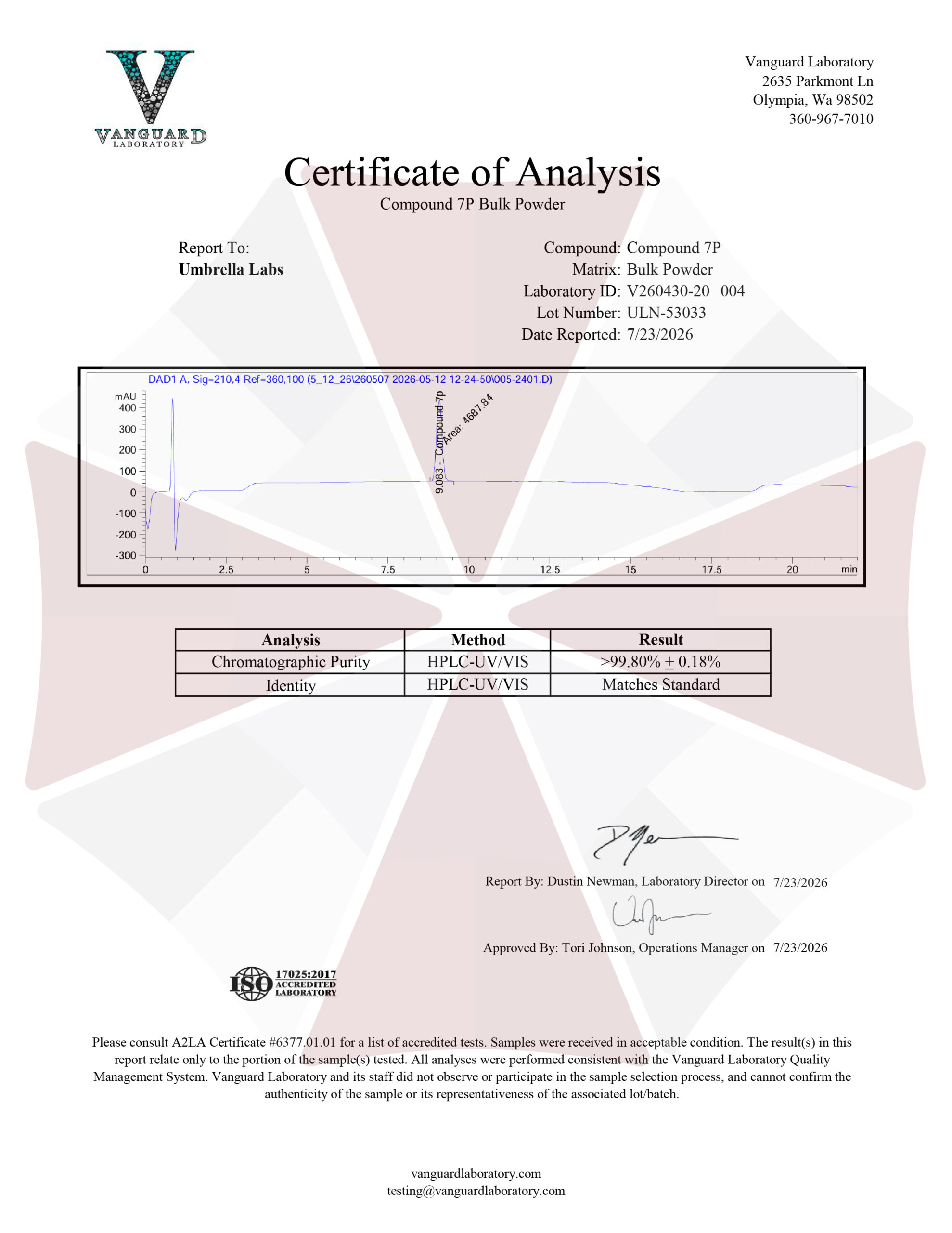
| File Name | View/Download |
| 2026-07-23-Umbrella-Labs-Compound-7P-Certificate-of-Analysis-COA.pdf |  |
VIEW CERTIFICATES OF ANALYSIS (COA)
Additional information
| Weight | 1 oz |
|---|---|
| Dimensions | 1.5 × 1.5 × 1.5 in |
| Options |