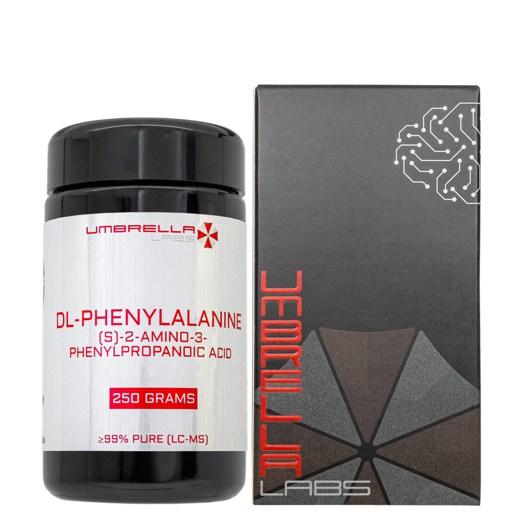
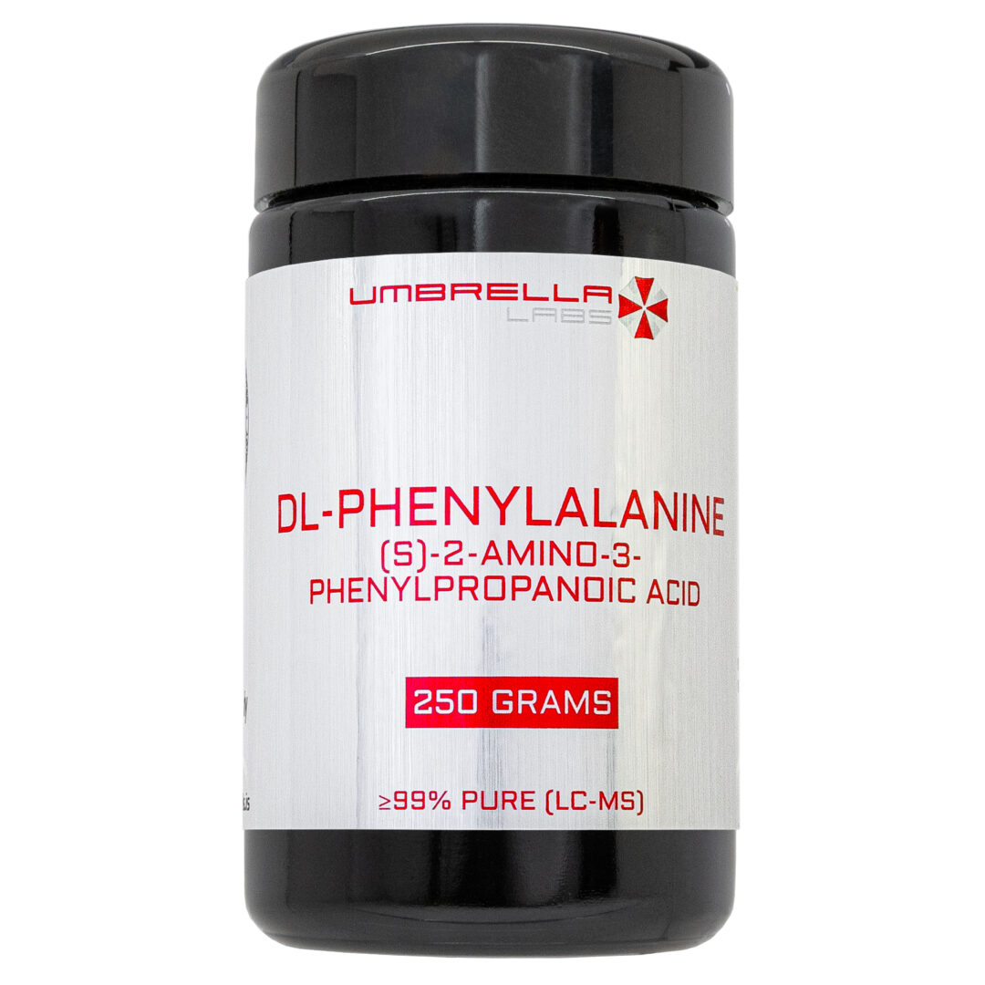
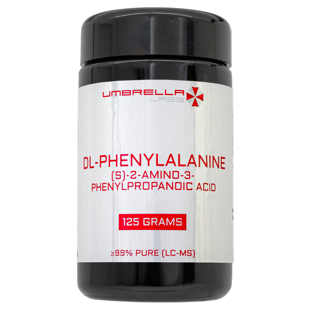
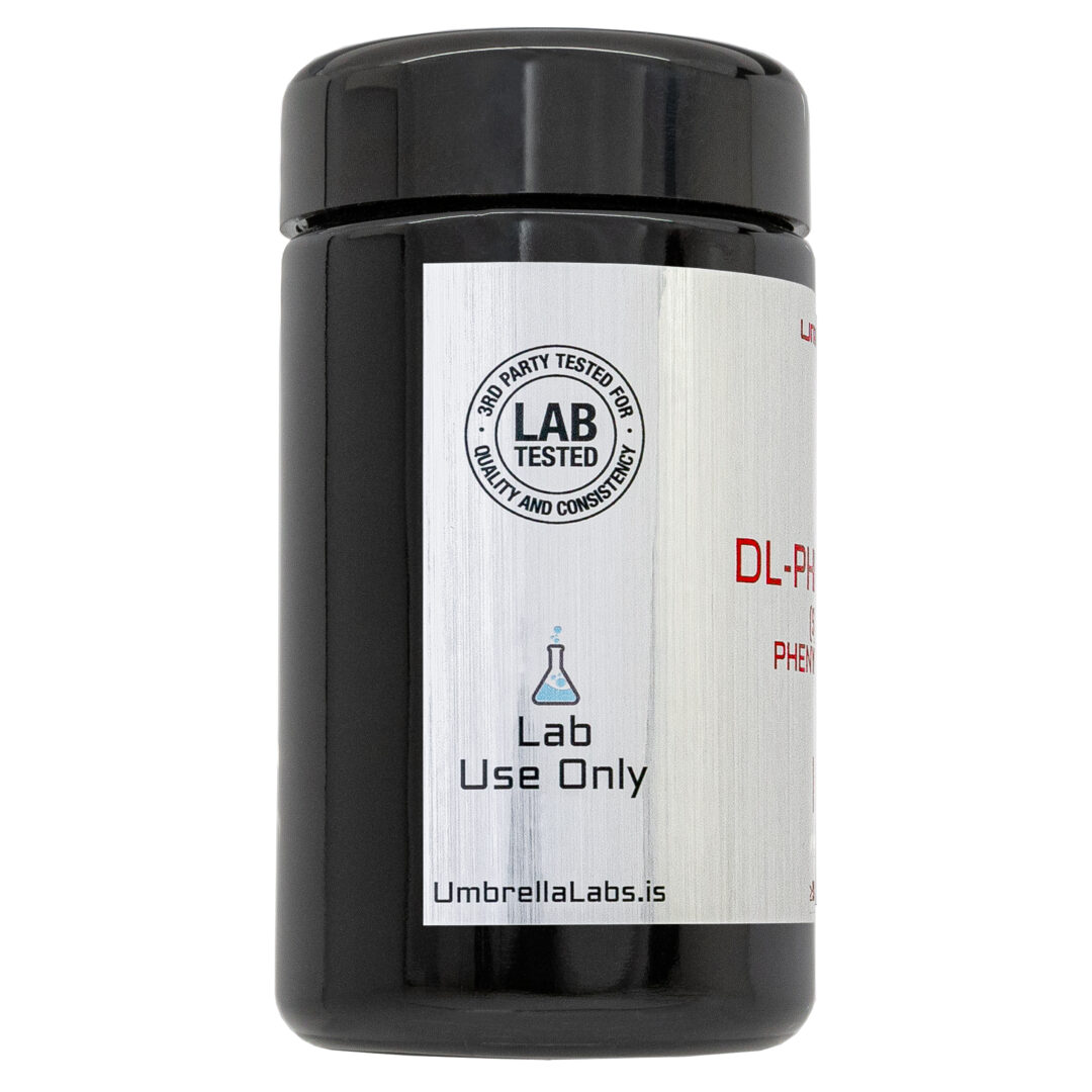
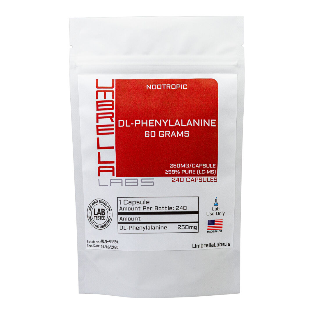
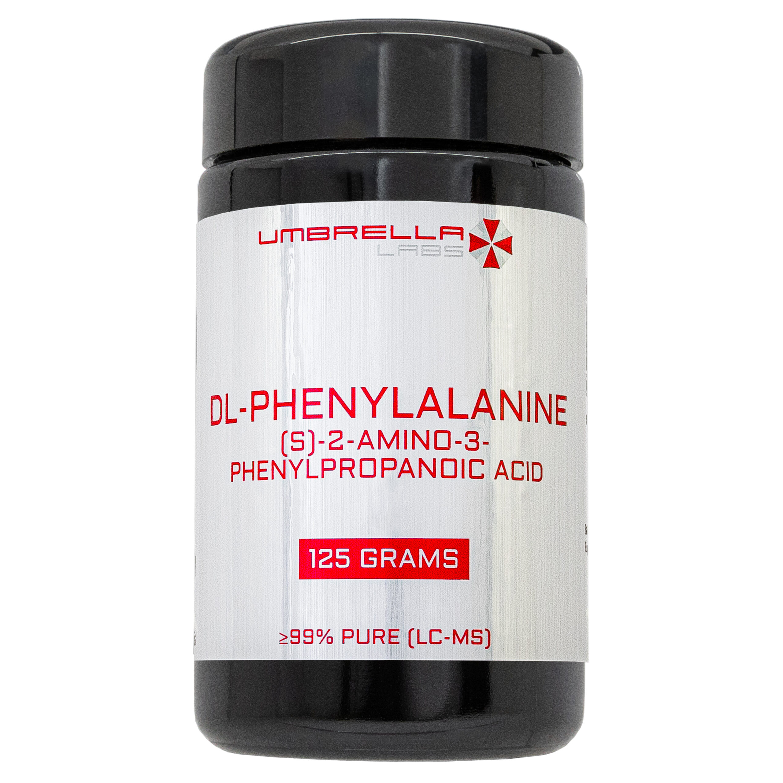
DL-PHENYLALANINE POWDER
$19.99 – $29.99Price range: $19.99 through $29.99
DL-Phenylalanine is sold for laboratory research use only. Terms of sale apply. Not for human consumption, nor medical, veterinary, or household uses. Please familiarize yourself with our Terms & Conditions prior to ordering.

- Description
- Additional information
Description
DL-Phenylalanine Nootropic Powder






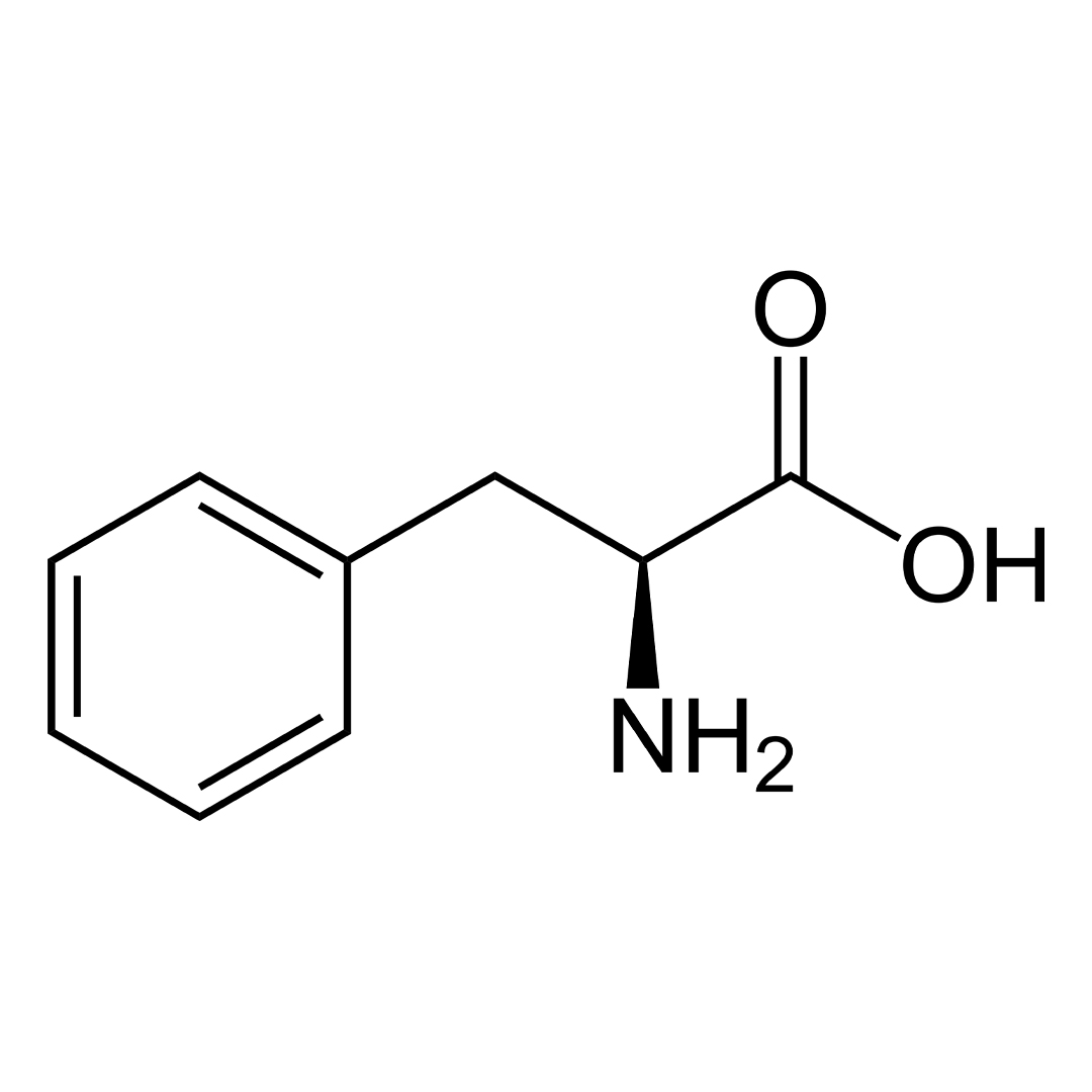
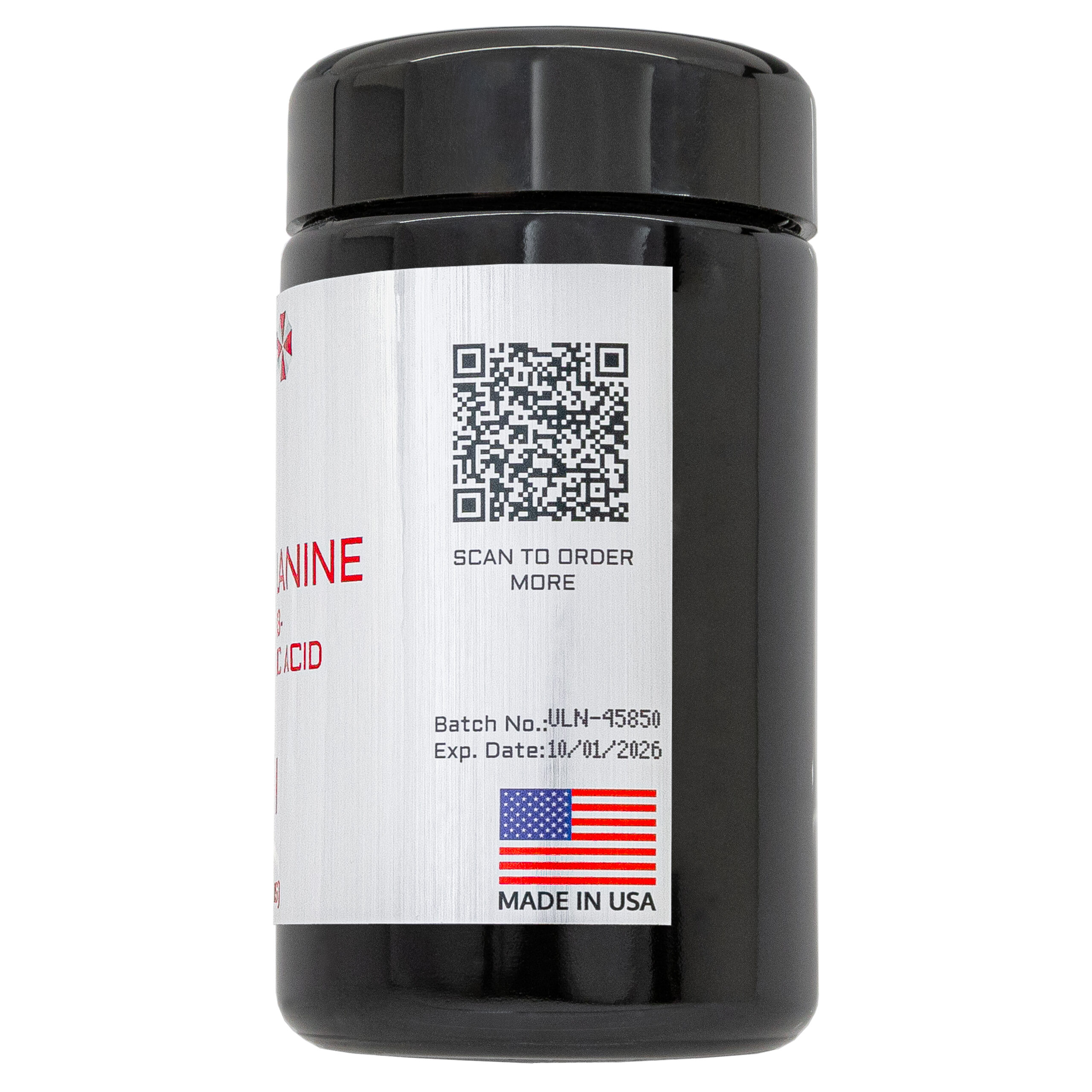
| CAS Number | 150-30-1 |
| Other Names | DL-3-Phenylalanine, H-DL-Phe-OH, DLPA, Phenylalanine DL-form |
| IUPAC Name | (S)-2-Amino-3-phenylpropanoic acid |
| Molecular Formula | C₉H₁₁NO₂ |
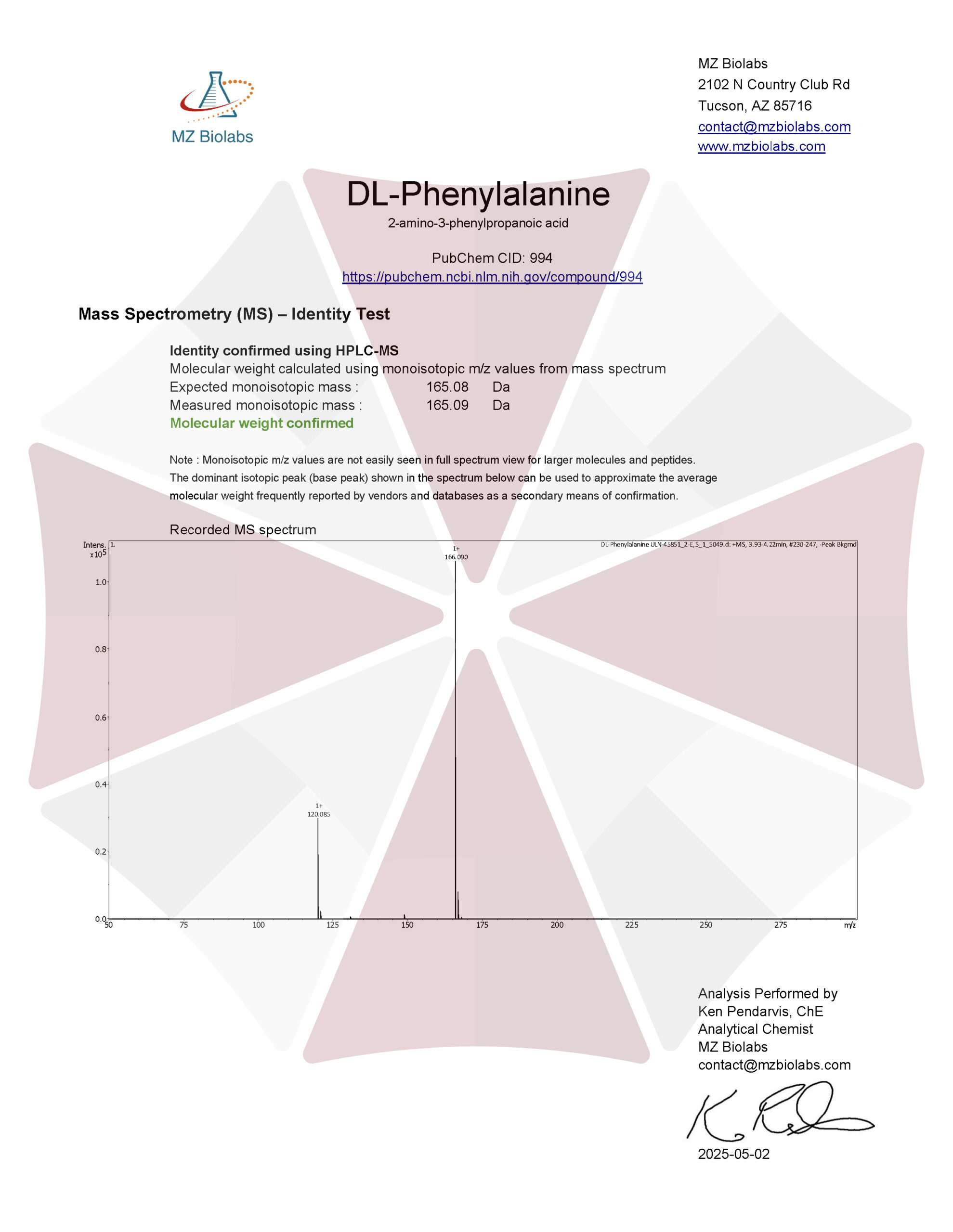
| Molecular Weight | 165.19 |
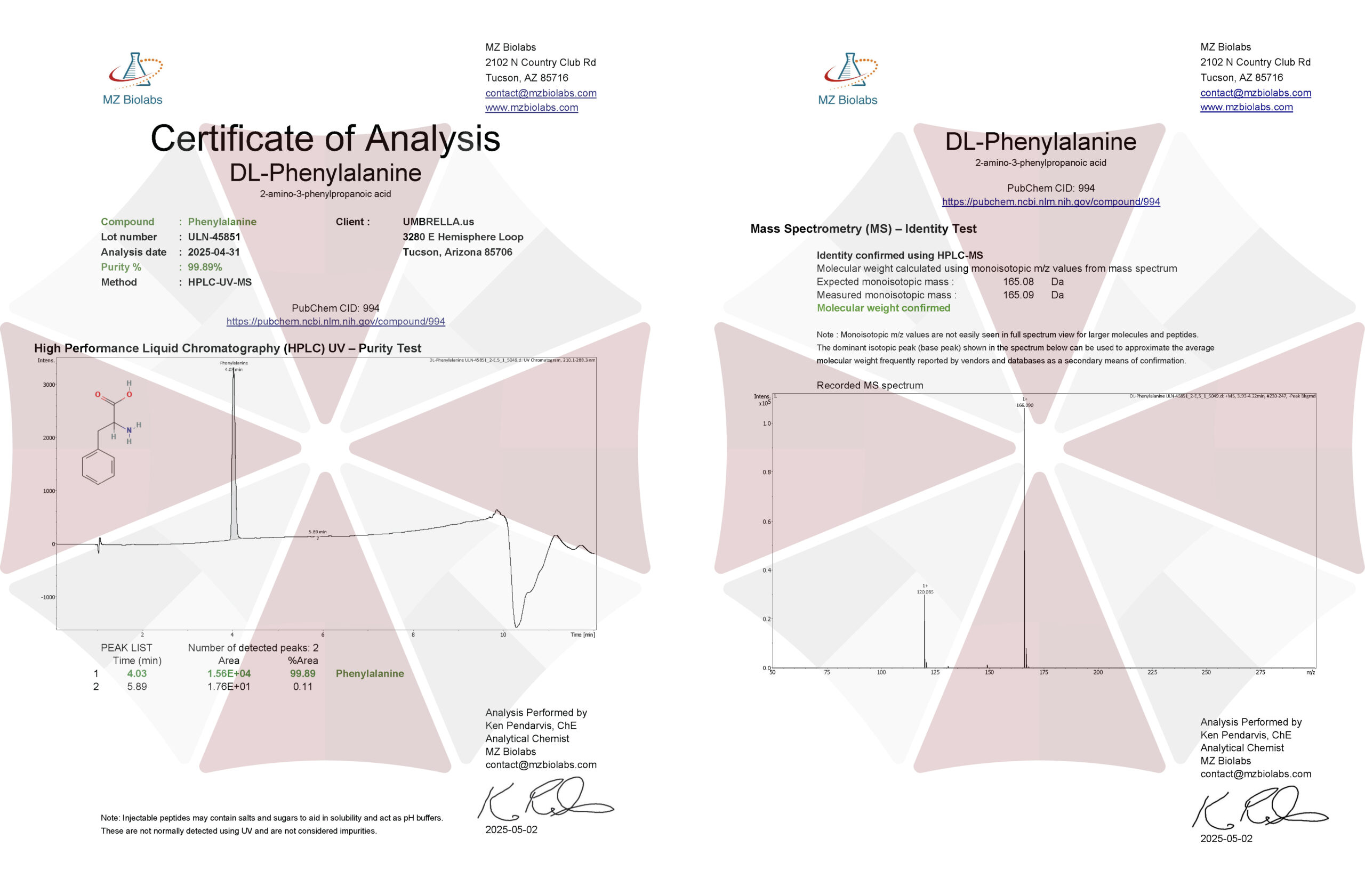
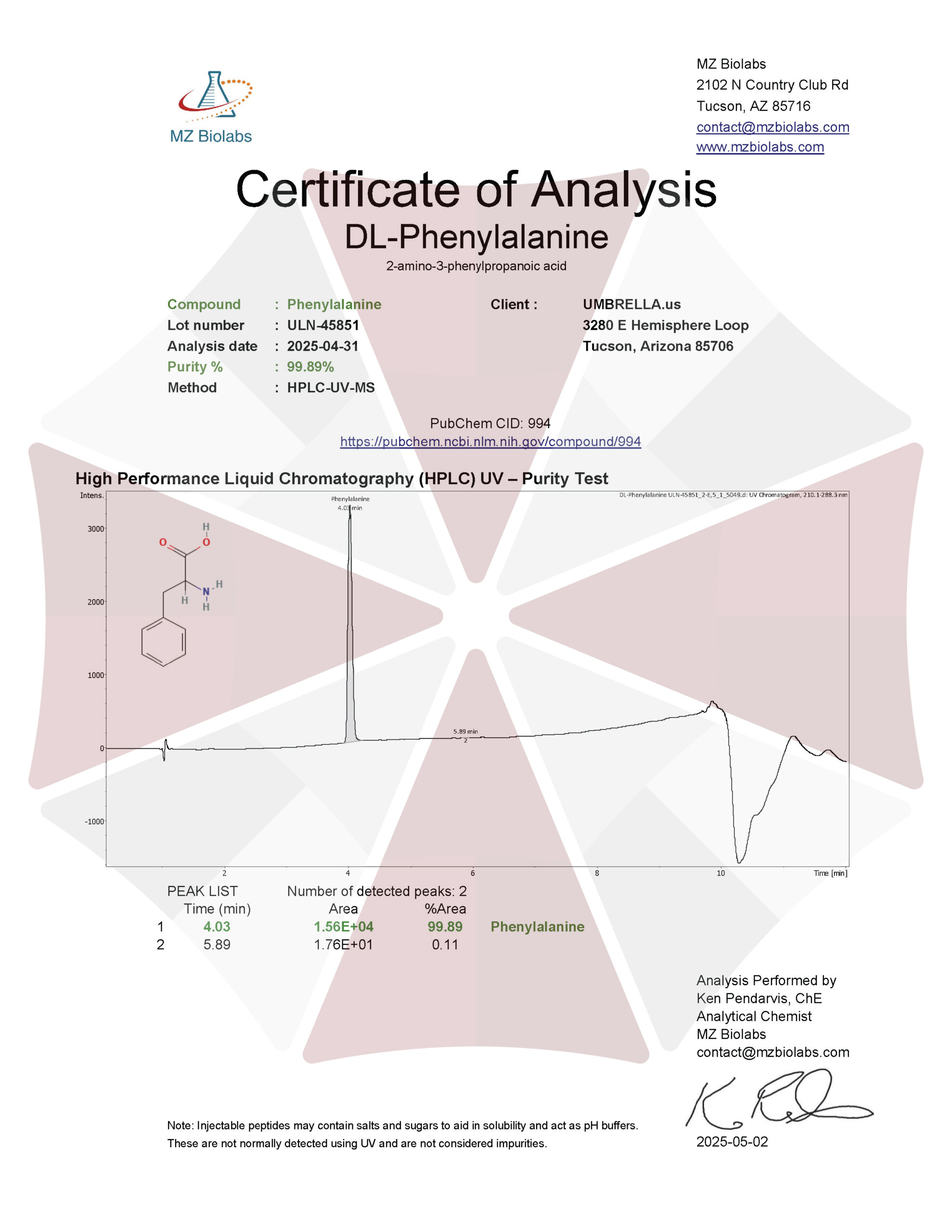
| Purity | ≥99% Pure (LC-MS) |
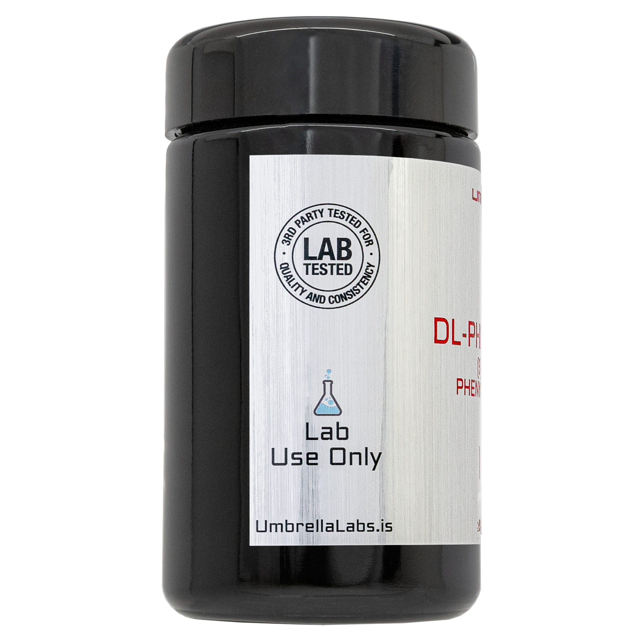
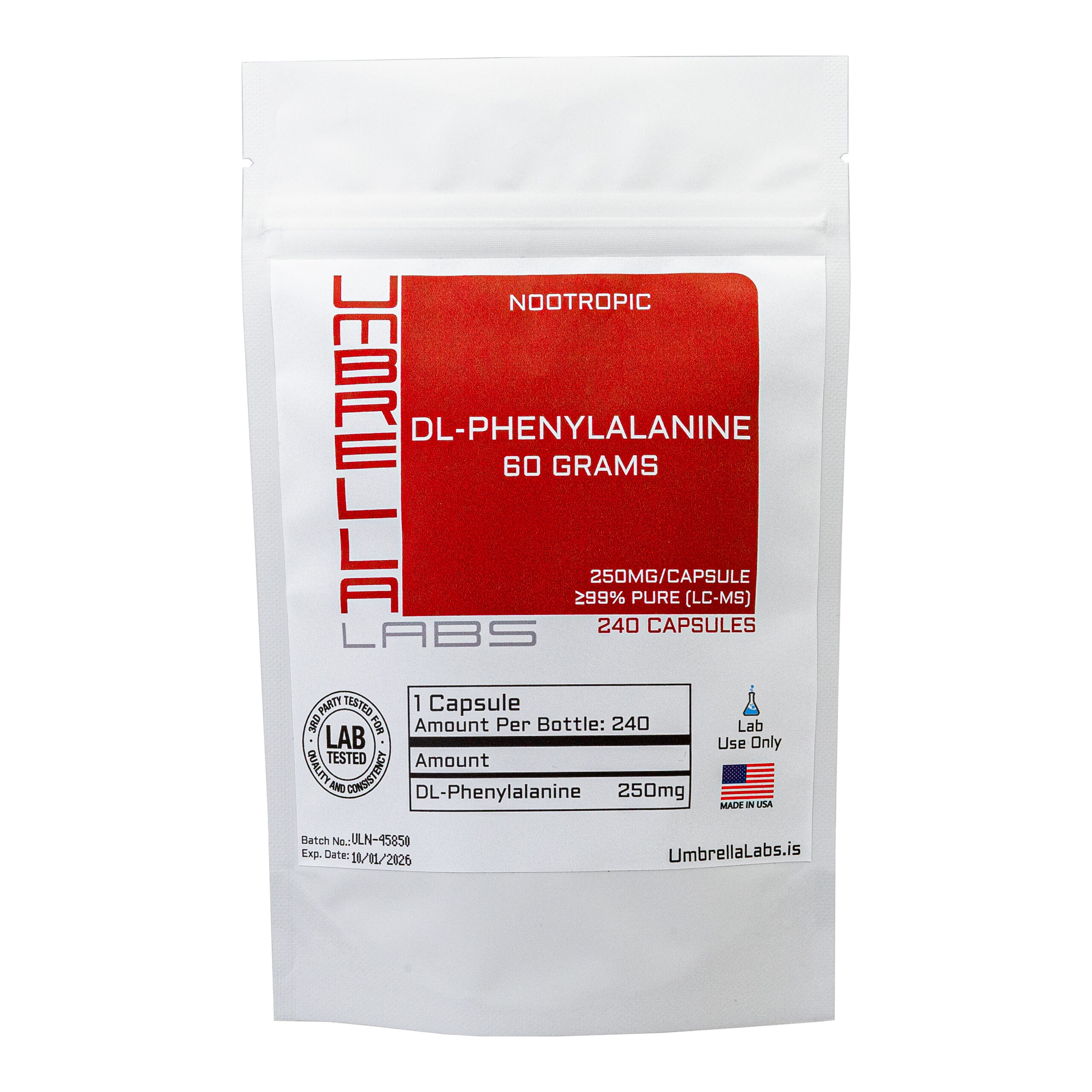
| Powder Availability |  125 grams, 250 grams, 240 capsules (250mg/capsule, 60 grams bottle) |
| Storage | Store in cool dry environment, away from direct sunlight. |
| Certificate of Analysis | Due to this product’s nature, this chemical does not have a COA associated with it. |
| Terms | All products are for laboratory developmental research USE ONLY. Products are not for human consumption. |
What is DL–Phenylalanine?
DL-Phenylalanine (DLPA) is a dietary amino acid supplement composed of equal parts L-phenylalanine, the naturally occurring essential amino acid, and D-phenylalanine, its synthetic mirror-image isomer. This unique combination has attracted interest as a potential nootropic due to its ability to support multiple neurochemical pathways involved in cognition, mood, and mental performance. L-phenylalanine serves as a precursor to the catecholamine neurotransmitters dopamine, norepinephrine, and epinephrine, which play critical roles in attention, motivation, learning, and executive function. D-phenylalanine, in contrast, has been investigated for its ability to inhibit the breakdown of endogenous endorphins and enkephalins, potentially enhancing mood, stress resilience, and overall mental well-being. Together, these complementary mechanisms have led to the exploration of DLPA as a cognitive enhancer capable of promoting alertness, reducing mental fatigue, and supporting emotional balance. Although preclinical and clinical studies suggest that DLPA may offer benefits for mood regulation, cognitive performance, and certain chronic pain conditions, the current body of evidence remains limited, and additional well-controlled human studies are needed to fully establish its efficacy, optimal dosing, and long-term safety as a nootropic supplement.
Main Research Findings
1) DL-phenylalanine was shown to protect against pulmonary vascular remodeling and lung inflammation induced by monocrotaline.
2) Concurrent treatment with DL-phenylalanine was found to potentiate pain relief and reduce depression in patients prescribed opiates for chronic pain management.
Selected Data
1) The study by the research team of Bai et al was designed to investigate whether 4-chloro-DL-phenylalanine (PCPA), a pharmacological inhibitor of tryptophan hydroxylase (Tph) and serotonin synthesis, could attenuate pulmonary vascular remodeling and lung inflammation in an experimental model of monocrotaline (MCT)-induced pulmonary arterial hypertension (PAH). A total of 68 male Sprague-Dawley rats weighing approximately 180 g were obtained from the Animal Resource Centre of China Medical University and maintained under controlled laboratory conditions with free access to food and water, a 12-hour light/dark cycle, ambient temperatures, and relative humidity of 50–70%. The animals were randomly assigned to four experimental groups consisting of an untreated control group, an MCT-treated group, an MCT plus low-dose PCPA group receiving 50 mg/kg/day, and an MCT plus high-dose PCPA group receiving 100 mg/kg/day. Pulmonary arterial hypertension was induced by a single intraperitoneal injection of monocrotaline at 60 mg/kg. Beginning immediately after MCT administration, rats assigned to the treatment groups received daily intraperitoneal injections of PCPA for 21 consecutive days, whereas the control and untreated MCT groups received equivalent volumes of physiological saline. Throughout the experiment, investigators performing all physiological, histological, and biochemical assessments remained blinded to treatment allocation to minimize experimental bias [1].
On day 22, the investigators evaluated the development of pulmonary hypertension using comprehensive hemodynamic measurements. Following anesthesia with pentobarbital sodium, systemic arterial pressure (SAP) was measured by catheterization of the right carotid artery, while pulmonary arterial pressure (PAP) was recorded by advancing a catheter through the right jugular vein into the pulmonary artery. Pressure signals were continuously monitored using pressure transducers connected to a multichannel polygraph recording system. After hemodynamic measurements were completed, animals were euthanized, and the hearts were dissected to separately weigh the right ventricle and the combined left ventricle plus interventricular septum. These measurements were used to calculate the right ventricular index as an indicator of right ventricular hypertrophy secondary to pulmonary hypertension. Body weight, heart rate, systemic arterial pressure, pulmonary arterial pressure, and ventricular hypertrophy were compared among all experimental groups to determine whether PCPA altered disease progression.
To evaluate structural changes within the pulmonary vasculature, lung tissues were perfusion-fixed, embedded in paraffin, sectioned at 5 μm, and stained with hematoxylin and eosin (H&E). Approximately twelve pulmonary arteries measuring 60–80 μm in external diameter were randomly selected from each animal for quantitative morphometric analysis using computerized image analysis software. Pulmonary vascular remodeling was assessed by calculating both medial wall thickness and wall area percentage, two established indices of smooth muscle hypertrophy and vascular wall thickening. Additional immunohistochemical analyses were performed on lung sections using antibodies directed against tryptophan hydroxylase-1 (Tph-1) and the serotonin transporter (SERT). Protein expression was visualized using diaminobenzidine staining, and staining intensity was quantified by measuring average optical density across multiple pulmonary arteries. These experiments were designed to determine whether inhibition of serotonin synthesis influenced the pulmonary expression of proteins central to serotonin production and transport [1].
The investigators next evaluated extracellular matrix remodeling and inflammatory signaling using several complementary molecular techniques. Gelatin zymography was performed on lung protein extracts to quantify the enzymatic activities of matrix metalloproteinase-2 (MMP-2) and MMP-9, two proteases involved in extracellular matrix degradation and vascular remodeling. Protein extracts were separated by sodium dodecyl sulfate-polyacrylamide gel electrophoresis using gelatin-containing gels, followed by incubation to allow proteolytic digestion and densitometric analysis of gelatinolytic activity.
In parallel, Western blot analysis was used to quantify protein expression of Tph-1, SERT, MMP-2, MMP-9, tissue inhibitors of metalloproteinases (TIMP-1 and TIMP-2), and the inflammatory mediators IL-1β, TNF-α, and intercellular adhesion molecule-1 (ICAM-1). Equal amounts of lung protein were separated by SDS-PAGE, transferred onto polyvinylidene difluoride membranes, incubated with specific primary and secondary antibodies, and visualized using enhanced chemiluminescence. Relative protein expression was quantified by densitometric analysis using β-actin as the loading control. Statistical analyses were performed using one-way analysis of variance followed by Fisher’s least significant difference or Dunnett’s T3 post hoc tests. This comprehensive methodological approach enabled the investigators to simultaneously evaluate the physiological, structural, biochemical, and inflammatory effects of PCPA treatment in experimental pulmonary arterial hypertension [1].
2) The research team of Russell et al developed their hypothesis by combining the senior author’s clinical experience treating patients with chronic non-malignant pain (CNMP) with an extensive review of previously published experimental and clinical literature concerning DL-phenylalanine (DLPA), endogenous opioid systems, serotonin signaling, and pain modulation. Rather than conducting a new randomized clinical trial or laboratory investigation, the paper synthesizes evidence from pharmacological, physiological, and clinical studies to explain why DLPA may enhance opioid analgesia when used as an adjunct to conventional pain management. The central objective was to propose a biological mechanism through which DLPA could improve pain relief, reduce depressive symptoms associated with chronic pain, and potentially permit reductions in opioid dosage while maintaining analgesic efficacy [2].
The clinical observations described in the article originated from the senior author’s treatment of more than 50 patients with chronic non-malignant pain using sustained-release opioid therapy, primarily long-acting morphine. During routine clinical management, patients who continued to experience incomplete pain control or depression despite opioid treatment were given supplemental DLPA. According to the author’s observations, DLPA was generally administered at doses of 500–1000 mg three times daily alongside morphine therapy. The physician monitored changes in pain relief, depressive symptoms, opioid requirements, and treatment tolerability over the course of clinical care. These observations served as the initial basis for developing the mechanistic hypothesis presented throughout the article.
To explain these observations, the authors performed a comprehensive review of the scientific literature describing the endogenous analgesia system (EAS). This review incorporated neuroanatomical, neurophysiological, pharmacological, and behavioral studies examining the descending pain-modulating pathways extending from the periaqueductal gray (PAG), nucleus raphe magnus (NRM), and nucleus reticularis paragigantocellularis (NRPG) to the dorsal horn of the spinal cord. The reviewed literature described how activation of these pathways suppresses transmission of nociceptive information through coordinated release of serotonin, norepinephrine, and endogenous opioid peptides such as enkephalins. The authors evaluated studies involving electrical brain stimulation, morphine administration, acupuncture, transcranial electrostimulation, transcutaneous electrical nerve stimulation (TENS), and cannabinoid-induced analgesia to illustrate the central role of the endogenous analgesia system in pain control [2].
The review further incorporated pharmacological studies examining the biological actions of D-phenylalanine, one component of DLPA. The authors analyzed evidence suggesting that D-phenylalanine inhibits enkephalinase, the enzyme responsible for degrading endogenous enkephalins, thereby prolonging opioid peptide activity within pain-modulating neural circuits. They also examined studies evaluating serotonin precursors such as tryptophan and 5-hydroxytryptophan, as well as serotonin reuptake inhibitors, because serotonin represents another major neurotransmitter within the endogenous analgesia system. By integrating findings from these diverse experimental models, the authors proposed that simultaneously enhancing both enkephalin and serotonin signaling could amplify endogenous analgesic mechanisms activated by opioids, acupuncture, cannabinoids, or other therapies.
Finally, the authors compared findings from animal experiments, human clinical trials, and observational studies evaluating D-phenylalanine or DLPA for chronic pain, depression, acupuncture analgesia, and opioid potentiation. They emphasized studies suggesting that D-phenylalanine enhanced the analgesic effects of morphine or acupuncture rather than functioning effectively as a stand-alone analgesic. Based on this synthesis of clinical observations and prior research, the paper proposed that future controlled clinical trials should formally evaluate combinations of DLPA with opioid therapy and potentially combine DLPA with other agents, including 5-hydroxytryptophan and serotonergic compounds, to maximize activation of the endogenous analgesia system while reducing opioid-related adverse effects [2].
Discussion
1) The study by Bai et al demonstrated that 4-chloro-DL-phenylalanine (PCPA) markedly attenuated the development of MCT-induced pulmonary arterial hypertension (PAH) by reducing pulmonary vascular remodeling, suppressing lung inflammation, and inhibiting serotonin-related signaling pathways. Over the three-week experimental period, MCT administration produced severe pulmonary hypertension accompanied by substantial mortality, with 8 of 17 rats in the untreated MCT group dying before study completion. In contrast, mortality was dramatically reduced in animals receiving PCPA, with only 1 of 17 rats dying in the low-dose treatment group. Hemodynamic measurements further confirmed the protective effects of PCPA. Compared with healthy control animals, MCT-treated rats developed significantly elevated PAP, pronounced right ventricular hypertrophy as measured by the right ventricular index, and marked reductions in body weight. Daily treatment with either 50 mg/kg or 100 mg/kg PCPA significantly lowered pulmonary arterial pressure and reduced right ventricular hypertrophy, although systemic arterial pressure and heart rate remained unchanged among all experimental groups. These findings demonstrated that inhibition of serotonin synthesis substantially improved the hemodynamic abnormalities associated with experimental pulmonary hypertension without affecting systemic cardiovascular function [1].

Figure 1: Changes in E) medial wall thickness of pulmonary arteries, and F) total wall area of pulmonary arteries.
Histological evaluation of lung tissues demonstrated that monocrotaline caused extensive pulmonary vascular remodeling characterized by thickening of the pulmonary arterial media and narrowing of the vascular lumen. Quantitative morphometric analysis revealed that medial wall thickness increased from approximately 19% in healthy control animals to nearly 50% following MCT administration, while vascular wall area also increased substantially, reflecting hypertrophy of pulmonary arterial smooth muscle. Both doses of PCPA significantly reduced these pathological changes, with the higher 100 mg/kg dose producing the greatest improvement. Representative histological images showed thinner vascular walls, larger luminal diameters, and an overall restoration of pulmonary arterial architecture in treated animals compared with untreated MCT rats. These structural improvements closely paralleled the observed reductions in pulmonary arterial pressure and right ventricular hypertrophy, indicating that PCPA effectively limited the vascular remodeling responsible for elevated pulmonary vascular resistance [1].
The investigators next examined serotonin-related molecular pathways and found that MCT-induced pulmonary hypertension markedly increased expression of both Tph-1, the rate-limiting enzyme responsible for peripheral serotonin synthesis, and SERT within pulmonary tissues. Immunohistochemical staining demonstrated intense Tph-1 and SERT immunoreactivity throughout pulmonary arteries in untreated MCT animals, while Western blot analysis confirmed significant increases in protein expression. Administration of PCPA reduced Tph-1 expression in a dose-dependent manner, with the higher dose producing near normalization of protein levels. SERT expression followed a similar pattern, with significant reductions observed after PCPA treatment using both immunohistochemistry and Western blot analysis. These findings indicated that pharmacological inhibition of tryptophan hydroxylase not only suppressed serotonin synthesis but also downregulated serotonin transporter expression, suggesting coordinated inhibition of the serotonin signaling pathway that has been implicated in pulmonary vascular remodeling and smooth muscle proliferation.
Further molecular analyses demonstrated that PCPA substantially inhibited extracellular matrix remodeling. Gelatin zymography revealed that monocrotaline markedly increased the enzymatic activities of MMP-2 and MMP-9, whereas treatment with PCPA reduced the activity of both enzymes, particularly at the higher dose. Western blot analyses produced similar results, showing significant MCT-induced increases in protein expression of MMP-2, MMP-9, TIMP-1, and TIMP-2, all of which were suppressed in a dose-dependent manner following PCPA administration. The reduction in both matrix metalloproteinases and their endogenous inhibitors suggested that PCPA restored extracellular matrix homeostasis rather than simply inhibiting one component of the remodeling process. These biochemical findings corresponded closely with the histological evidence of reduced vascular wall thickening, indicating that suppression of matrix remodeling contributed substantially to the structural protection afforded by PCPA [1].
The study also demonstrated pronounced anti-inflammatory effects of PCPA. Histological examination revealed extensive perivascular inflammatory cell infiltration in untreated MCT animals, whereas both doses of PCPA markedly reduced inflammatory cell accumulation within lung tissue. Consistent with these observations, Western blot analyses showed significant MCT-induced increases in the pro-inflammatory cytokines IL-1β, TNF-α, and ICAM-1. Treatment with PCPA significantly reduced expression of all three inflammatory mediators, with the higher dose producing the greatest suppression. Collectively, the results demonstrated that inhibition of serotonin synthesis by PCPA simultaneously reduced pulmonary hypertension, right ventricular hypertrophy, vascular remodeling, extracellular matrix turnover, and inflammatory signaling. The authors concluded that the protective effects of PCPA were mediated through coordinated downregulation of Tph-1, SERT, MMP/TIMP signaling, and multiple inflammatory cytokines, thereby limiting both structural and inflammatory processes that contribute to the progression of monocrotaline-induced pulmonary arterial hypertension [1].

Figure 2: Western blot analysis representing changes in levels of A) IL-1β, B) TNF-α, and C) ICAM-1.
2) The research team of Russel et al report that, in their clinical experience, DLPA consistently enhanced the therapeutic effects of long-acting morphine in patients with CNMP. Patients receiving DLPA in combination with sustained-release morphine frequently experienced improved pain control, better mood, and, in many cases, sufficient improvement to permit reductions in morphine dosage. The authors emphasize that these observations were made during routine clinical practice rather than through a controlled clinical trial. The consistency of these observations prompted the authors to explore possible neurobiological mechanisms that could explain the apparent synergistic interaction between DLPA and opioid analgesics [2].
A major conclusion of the paper is that DLPA likely exerts its beneficial effects by enhancing activity within the EAS. The authors review extensive neurophysiological evidence demonstrating that the EAS functions as a descending inhibitory pathway originating in the periaqueductal gray, nucleus raphe magnus, and related brainstem nuclei. Activation of this system suppresses pain transmission within the dorsal horn of the spinal cord through the coordinated release of serotonin, norepinephrine, and endogenous opioid peptides, particularly enkephalins. Morphine, acupuncture, transcranial electrostimulation, and several other analgesic interventions all activate this pathway. The authors propose that DLPA augments these naturally occurring analgesic mechanisms by prolonging the activity of endogenous enkephalins through inhibition of enkephalinase while simultaneously supporting neurotransmitter systems involved in descending pain inhibition. According to this hypothesis, DLPA does not replace opioid therapy but instead amplifies the body’s intrinsic pain-modulating pathways, thereby increasing the effectiveness of conventional analgesics.
The literature reviewed by the authors also provides indirect support for this proposed mechanism. Animal studies cited throughout the paper demonstrated that D-phenylalanine enhanced morphine-induced analgesia, allowing equivalent pain relief to be achieved with lower morphine doses. Other experimental investigations showed that D-phenylalanine potentiated analgesia produced by acupuncture, stress-induced analgesia, transcranial electrical stimulation, cannabinoids, and several antidepressant medications. Similar enhancement was observed with serotonin precursors such as L-tryptophan and 5-hydroxytryptophan, as well as serotonin reuptake inhibitors, supporting the concept that serotonin and endogenous opioid peptides function cooperatively within the endogenous analgesia system. The authors therefore conclude that simultaneous enhancement of serotonergic and enkephalinergic neurotransmission represents a rational strategy for strengthening endogenous pain inhibition and improving clinical analgesia [2].
The review also evaluated previous clinical investigations of D-phenylalanine and DLPA. While the authors acknowledged that studies examining D-phenylalanine as a stand-alone analgesic produced inconsistent findings, including one double-blind clinical trial that failed to demonstrate significant benefit for chronic pain, they emphasized that most positive findings involved combination therapy rather than monotherapy. Reports showing enhanced acupuncture analgesia and improved morphine efficacy suggested that D-phenylalanine functions primarily as an adjunctive treatment capable of potentiating existing analgesic interventions instead of acting independently. Likewise, earlier studies evaluating DLPA for depression produced mixed outcomes, although one double-blind trial found antidepressant effects comparable to imipramine. The authors suggest that improvements in mood observed among patients with chronic pain may have resulted both from direct neurochemical effects and from better pain control achieved through enhanced endogenous analgesia.
Based on the combined clinical observations and literature review, the authors conclude that DLPA represents a promising adjunct to chronic opioid therapy rather than a replacement for established analgesic treatments. They propose that supplementation with DLPA may improve analgesic efficacy, reduce opioid dosage requirements, lessen opioid-associated adverse effects such as sedation and constipation, and simultaneously alleviate the depression frequently accompanying chronic pain. The paper further suggests that combining DLPA with other agents that enhance serotonin activity, including 5-hydroxytryptophan, St. John’s wort, and potentially tyrosine, may provide even greater activation of the endogenous analgesia system. However, the authors repeatedly emphasize that these conclusions remain hypothetical and must be confirmed through well-designed randomized, placebo-controlled clinical trials specifically evaluating DLPA as an adjunctive therapy for chronic non-malignant pain [2].
Disclaimer
**LAB USE ONLY**
*This information is for educational purposes only and does not constitute medical advice. THE PRODUCTS DESCRIBED HEREIN ARE FOR RESEARCH USE ONLY. All clinical research must be conducted with oversight from the appropriate Institutional Review Board (IRB). All preclinical research must be conducted with oversight from the appropriate Institutional Animal Care and Use Committee (IACUC) following the guidelines of the Animal Welfare Act (AWA).
Citations
[1] Bai Y, Wang HM, Liu M, et al. 4-Chloro-DL-phenylalanine protects against monocrotaline‑induced pulmonary vascular remodeling and lung inflammation. Int J Mol Med. 2014;33(2):373-382. doi:10.3892/ijmm.2013.1591
[2] Russell AL, McCarty MF. DL-phenylalanine markedly potentiates opiate analgesia – an example of nutrient/pharmaceutical up-regulation of the endogenous analgesia system. Med Hypotheses. 2000;55(4):283-288. doi:10.1054/mehy.1999.1031
DL-Phenylalanine is sold for laboratory research use only. Terms of sale apply. Not for human consumption, nor medical, veterinary, or household uses. Please familiarize yourself with our Terms & Conditions before ordering.
| File Name | View/Download |
| 2025-04-31-Umbrella-Labs-DL-Phenylalanine-Certificate-Of-Analysis-COA.pdf |  |
VIEW CERTIFICATES OF ANALYSIS (COA)
Additional information
| Weight | N/A |
|---|---|
| Dimensions | N/A |
| Options |