HBT1 (YDL223C) POWDER (1 GRAM)
$79.99
HBT1 (YDL223C) is sold for laboratory research use only. Terms of sale apply. Not for human consumption, nor medical, veterinary, or household uses. Please familiarize yourself with our Terms & Conditions prior to ordering.

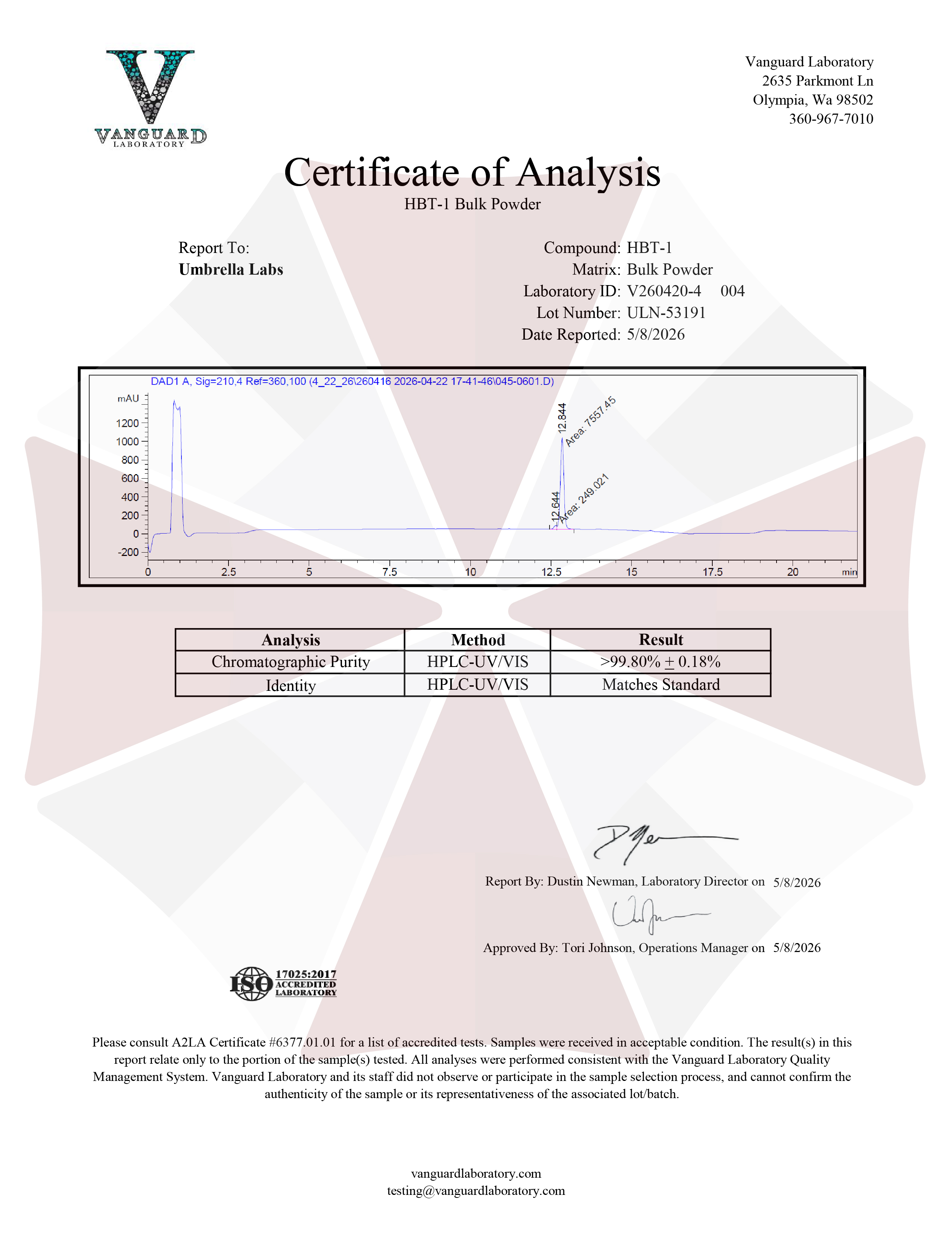
Also Available In:
 Liquid Option >>
- Description
- Additional information
Description
HBT1 (YDL223C) Nootropic Powder


| CAS Number | 489408-02-8 |
| Other Names | AKOS024270212, BS-47734, 2-[[2-[5-Methyl-3-(trifluoromethyl)pyrazol-1-yl]acetyl]amino]-4,5,6,7-tetrahydro-1-benzothiophene-3-carboxamide |
| IUPAC Name | 2-[[2-[5-methyl-3-(trifluoromethyl)pyrazol-1-yl]acetyl]amino]-4,5,6,7-tetrahydro-1-benzothiophene-3-carboxamide |
| Molecular Formula | C₁₆H₁₇F₃N₄O₂S |
| Molecular Weight | 386.39 |
| Purity | ≥99% Pure (LC-MS) |
| Liquid Availability | 30mL liquid (20mg/mL, 600mg bottle) |
| Powder Availability | 1 gram |
| Storage | Store in cool dry environment, away from direct sunlight. |
| Terms | All products are for laboratory developmental research USE ONLY. Products are not for human consumption. |
What is HBT1?
HBT1 is an experimental nootropic compound that has attracted interest for its potential to enhance cognitive function through selective modulation of glutamatergic neurotransmission. Unlike conventional glutamate receptor agonists, HBT1 functions as a positive allosteric modulator (PAM) of AMPA receptors, enhancing receptor activity only in the presence of the endogenous neurotransmitter glutamate rather than directly activating the receptor itself. This mechanism is intended to strengthen synaptic plasticity while minimizing the risk of excessive neuronal excitation associated with direct agonists. Preclinical studies have demonstrated that HBT1 enhances long-term potentiation (LTP), a cellular process underlying learning and memory, and improves performance in animal models of cognitive impairment without producing significant adverse neurological effects. Because of its favorable pharmacological profile and ability to facilitate excitatory neurotransmission in a controlled manner, HBT1 has emerged as a promising candidate for the treatment of cognitive deficits associated with aging, neurodegenerative diseases such as Alzheimer’s disease, and other disorders characterized by impaired synaptic function. HBT1 represents a novel approach to cognitive enhancement by targeting the mechanisms responsible for memory formation and neural plasticity rather than simply increasing neurotransmitter release.
Main Research Findings
1) HBT1 was shown to avoid a bell-shaped curve response in in vitro BDNFproduction.
2) Use of AMPA receptors similar to HBT1 was found to increase the cortical excitability threshold in patients with amyotrophic lateral sclerosis.
Selected Data
1) The study performed by Kunugi et al employed a comprehensive series of in vitro pharmacological, electrophysiological, biochemical, molecular, and structural biology techniques to identify and characterize HBT1, a novel AMPA receptor (AMPA-R) positive allosteric modulator with reduced agonistic activity compared with previously developed AMPA-R potentiators. The investigators aimed to determine whether minimizing agonistic activity could reduce the bell-shaped dose-response relationship commonly observed with other AMPA receptor modulators while maintaining their ability to enhance brain-derived neurotrophic factor (BDNF) production [1].
Primary hippocampal neurons were prepared from Sprague-Dawley rat embryos at embryonic day 19. Following dissection of the fetal hippocampi, neuronal tissue was enzymatically dissociated and plated onto poly-D-lysine- or poly-L-lysine-coated culture plates depending on the experimental assay. Cells were maintained in Neurobasal medium supplemented with B27, L-glutamine, penicillin, streptomycin, and gentamicin under standard cell culture conditions until they reached the appropriate maturation stage for each experiment. These primary neuronal cultures served as the principal experimental model because they more closely resemble native neuronal AMPA receptor physiology than recombinant cell lines.
To assess neurotrophic signaling, cultured hippocampal neurons were treated for 24 hours with HBT1, OXP1, LY451395, or LY451646 in either the presence or absence of low concentrations of AMPA. After treatment, cells were lysed and intracellular BDNF protein concentrations were quantified using a commercially available enzyme-linked immunosorbent immunoassay. Because excessive AMPA receptor activation can produce excitotoxicity, the investigators simultaneously evaluated cellular toxicity using a lactate dehydrogenase (LDH) release assay, in which extracellular LDH activity served as an indicator of membrane damage and neuronal death. These complementary assays allowed the researchers to determine whether changes in BDNF production reflected pharmacological activity rather than cytotoxic effects [1].
Electrophysiological characterization was performed using whole-cell patch-clamp recordings in cultured hippocampal neurons between 11 and 20 days after plating. Neurons were voltage-clamped while inward AMPA receptor-mediated currents were recorded before and after application of the test compounds, both in the presence and absence of exogenous AMPA. These experiments directly measured whether each compound functioned solely as a positive allosteric modulator or possessed intrinsic agonistic properties capable of activating AMPA receptors independently of glutamate.
The investigators also performed calcium influx assays using fluorescent calcium indicator dyes in both primary neurons and recombinant Chinese hamster ovary (CHO) cell lines expressing individual human AMPA receptor subunits together with transmembrane AMPA receptor regulatory proteins (TARPs). Calcium responses were measured after exposure to the test compounds with or without glutamate or AMPA. Additional experiments evaluated receptor subtype selectivity by testing multiple GluA1–GluA4 splice variants and examined potential off-target activity at kainate receptors. Pharmacological specificity was further confirmed through experiments using the competitive AMPA receptor antagonist NBQX. These assays enabled comparison between native neuronal receptors and recombinant receptor systems while determining whether HBT1 required endogenous glutamate for receptor activation [1].
To investigate receptor binding mechanisms, the researchers generated recombinant GluA2 ligand-binding domain (LBD) and amino-terminal domain (ATD) proteins. Radiolabeled forms of HBT1 and OXP1 were synthesized and used in scintillation proximity assays (SPA) to quantify ligand binding affinity, determine glutamate dependence, and identify competitive interactions between compounds. Additional membrane binding experiments were conducted using rat hippocampal tissue to evaluate interactions with native AMPA receptors. Competitive displacement assays with radiolabeled AMPA determined whether HBT1 or related compounds occupied the orthosteric glutamate-binding site or acted through distinct allosteric binding sites [1].
Structural characterization was performed using X-ray crystallography of purified GluA2 ligand-binding domains co-crystallized with glutamate and either HBT1 or LY451395. Recombinant proteins were expressed, purified through affinity and size-exclusion chromatography, crystallized using vapor diffusion techniques, and analyzed by synchrotron X-ray diffraction. Molecular replacement and iterative refinement generated high-resolution structures that identified ligand binding positions and amino acid interactions responsible for receptor modulation. These structural analyses allowed direct comparison of the molecular binding modes of HBT1 and LY451395 within the AMPA receptor ligand-binding domain.
Finally, all experimental data were analyzed using appropriate statistical procedures, including Student’s t-tests, Aspin-Welch tests, Bartlett’s tests, Williams’ tests, Shirley-Williams tests, and Dunnett’s multiple-comparison tests, depending on the experimental design and distribution of the data. Concentration-response relationships and EC₅₀ values were calculated using logistic regression analysis, with statistical significance established according to predefined probability thresholds. Collectively, this multidisciplinary experimental approach integrated cellular pharmacology, electrophysiology, receptor pharmacology, structural biology, and quantitative statistical analyses to comprehensively evaluate HBT1 as a novel AMPA receptor potentiator with reduced agonistic properties and improved pharmacological characteristics [1].
2) The study by the research team of Oskarsson et al was designed as a randomized, double-blind, placebo-controlled, single-center pilot clinical trial to determine whether the AMPA receptor antagonist perampanel could alter cortical excitability in patients with ALS. The investigation sought to establish whether transcranial magnetic stimulation (TMS) could serve as a functional biomarker of target engagement following acute AMPA receptor blockade. Because cortical hyperexcitability is a recognized feature of ALS and is believed to result largely from excessive glutamatergic signaling through AMPA receptors, the investigators hypothesized that pharmacologic inhibition of these receptors would increase the resting (MT), thereby providing physiological evidence that perampanel reached its intended central nervous system target [2].
Eligible participants were adults 18 to 70 years of age with clinically probable laboratory-supported, clinically probable, or clinically definite ALS according to the revised El Escorial diagnostic criteria. To ensure reliable electrophysiological measurements, subjects were required to retain sufficient strength in the abductor pollicis brevis muscle to overcome gravity, as this muscle served as the recording site for motor evoked potentials. Before randomization, each participant underwent baseline TMS testing to establish a measurable resting motor threshold. Individuals in whom no threshold could be identified were excluded from participation. Additional exclusion criteria included a history of epilepsy, skull defects, other contraindications to TMS, aggressive behavior, substance abuse, or unwillingness to abstain from alcohol for two weeks following dosing. These eligibility criteria were intended to maximize participant safety while ensuring high-quality neurophysiological recordings.
After successful screening, participants were randomly assigned in a 5:1 ratio to receive either oral perampanel or matching placebo. During the initial treatment phase, participants received a single 4-mg dose of perampanel or placebo. More than three weeks later, returning participants underwent a second treatment period in which they received 8 mg of perampanel or placebo. Although the investigators originally planned to enroll twelve participants in the second-dose cohort, the trial was prematurely terminated because of COVID-19-related research restrictions after only seven individuals completed the higher-dose phase. Throughout each dosing session, participants underwent repeated physiological assessments for four hours after administration and received follow-up telephone evaluations the following day to identify delayed adverse events [2].
Cortical excitability was measured using transcranial magnetic stimulation performed by a board-certified electrodiagnostic specialist. A magnetic stimulator equipped with a figure-of-eight coil was positioned over the primary motor cortex to identify the optimal stimulation site capable of eliciting motor evoked potentials from the abductor pollicis brevis muscle and surface electromyographic recordings were obtained. The resting motor threshold was defined as the minimum stimulation intensity capable of producing motor evoked potentials greater than 50 μV in at least five of ten trials, consistent with recommendations from the International Federation of Clinical Neurophysiology. To ensure consistent coil positioning throughout repeated measurements, participants wore a marked, tight-fitting skull cap. Motor threshold measurements were obtained immediately before drug administration and then repeated hourly for four hours after dosing. In addition to the primary resting motor threshold measurement, investigators also calculated lower and upper motor thresholds to provide complementary measures of cortical excitability [2].
Blood samples were collected hourly during the four-hour observation period to determine serum perampanel concentrations, enabling investigators to compare pharmacokinetic profiles with physiological changes in cortical excitability. Safety assessments included monitoring adverse events during the study visit and during the follow-up telephone interview. Disease severity was documented using the revised Amyotrophic Lateral Sclerosis Functional Rating Scale (ALSFRS-R). Continuous variables were summarized using medians and interquartile ranges, whereas categorical variables were presented as frequencies and percentages. Because of the small sample size, changes in motor threshold, lower threshold, and upper threshold from baseline to two hours after dosing were analyzed using the Wilcoxon signed-rank test, with two-sided P values below 0.05 considered statistically significant. When motor thresholds became unobtainable during testing, stimulation intensity values were assigned the maximum value of 100% of stimulator output to permit statistical analysis. This comprehensive methodology combined standardized neurophysiological measurements, pharmacokinetic monitoring, clinical assessments, and appropriate nonparametric statistical analyses to determine whether acute AMPA receptor blockade could measurably alter cortical excitability in ALS patients [2].
Discussion
1) The study by Kunugi et al demonstrated that HBT1 possesses a distinct pharmacological profile compared with previously developed AMPA receptor potentiators by enhancing AMPA receptor function while exhibiting minimal intrinsic agonistic activity. The investigators first confirmed that the established AMPA receptor potentiators LY451646 and LY451395 displayed undesirable bell-shaped dose-response curves for BDNF production in cultured rat hippocampal neurons. Both compounds significantly increased BDNF production at low to moderate concentrations but lost efficacy as concentrations increased. Importantly, these compounds also stimulated BDNF production even in the absence of exogenous AMPA, indicating intrinsic agonistic activity rather than purely positive allosteric modulation. Lactate dehydrogenase release assays demonstrated that these bell-shaped responses were not primarily caused by cytotoxicity, suggesting that excessive receptor activation contributed to the loss of efficacy at higher concentrations. These findings led the authors to propose that intrinsic agonistic activity may underlie the bell-shaped pharmacological responses observed with earlier AMPA receptor potentiators [1].
Electrophysiological studies supported this hypothesis by showing that LY451395 generated AMPA receptor-mediated currents even in the absence of agonist during whole-cell patch-clamp recordings from primary hippocampal neurons. Calcium influx experiments further demonstrated that LY451395 produced calcium entry in primary neurons both with and without AMPA. Interestingly, this agonistic behavior was absent in recombinant CHO cells expressing AMPA receptors, where receptor activation required glutamate. These findings revealed an important difference between native neuronal AMPA receptors and recombinant receptor systems, indicating that conventional screening methods using transfected cell lines may fail to identify compounds with undesirable agonistic properties. Competitive binding experiments also demonstrated that LY451395 did not displace radiolabeled AMPA from its orthosteric binding site, indicating that receptor activation occurred through allosteric rather than direct agonist binding [1].
Screening of the compound library identified two novel AMPA receptor potentiators, HBT1 and OXP1, which were subsequently characterized in detail. Both compounds potentiated AMPA receptor-mediated calcium influx in recombinant cells and primary neurons in a glutamate-dependent manner, confirming that they functioned as positive allosteric modulators. However, significant differences emerged during electrophysiological testing. Whole-cell patch-clamp recordings showed that HBT1 produced receptor-mediated currents only when AMPA was present, whereas OXP1 activated AMPA receptors even in the absence of agonist, resembling the behavior of LY451395. These observations demonstrated that HBT1 exhibited substantially lower intrinsic agonistic activity than either OXP1 or previously developed AMPA receptor potentiators.
The BDNF production assays further highlighted the advantages of HBT1. In the presence of AMPA, HBT1 produced a concentration-dependent increase in BDNF protein that remained elevated across a broad concentration range without displaying the bell-shaped decline observed for LY451646, LY451395, and OXP1. Furthermore, HBT1 failed to stimulate BDNF production in the absence of AMPA at therapeutically relevant concentrations, confirming its low agonistic profile. By contrast, OXP1 displayed both agonistic activity and a bell-shaped dose-response similar to the earlier compounds. Cytotoxicity measurements also favored HBT1, as it produced no significant lactate dehydrogenase release either in the presence or absence of AMPA, whereas OXP1 caused modest but statistically significant toxicity at higher concentrations. These results strongly suggested that reducing intrinsic agonistic activity decreases the likelihood of bell-shaped pharmacological responses while preserving neurotrophic efficacy [1].
Binding studies revealed that HBT1 interacted directly with the ligand-binding domain (LBD) of the AMPA receptor in a glutamate-dependent manner. Radiolabeled HBT1 bound specifically to recombinant GluA2 ligand-binding domains and to native rat hippocampal membranes only when glutamate was present, indicating that receptor activation required endogenous agonist binding. Structural analysis using X-ray crystallography showed that HBT1 occupied the established allosteric pocket at the ligand-binding domain dimer interface but adopted a binding orientation distinct from LY451395. Unlike LY451395, HBT1 formed hydrogen bonds with the amino acid residue Ser518, providing a unique interaction that may contribute to its reduced agonistic activity. In contrast, OXP1 did not bind detectably to the same ligand-binding pocket and instead appeared to interact with a separate receptor site, possibly involving the amino-terminal domain, although its precise binding location could not be definitively established.
Additional experiments demonstrated that OXP1 enhanced HBT1 binding to AMPA receptors and that simultaneous application of both compounds activated receptors even in the absence of agonist, further supporting the existence of multiple allosteric binding sites capable of cooperatively modulating receptor activity. Collectively, the results showed that HBT1 functions as a glutamate-dependent AMPA receptor potentiator with minimal intrinsic agonistic activity, sustained enhancement of BDNF production across a wide concentration range, low cytotoxicity, and a unique structural binding mode. Although the authors noted that HBT1 itself possesses poor pharmacokinetic properties that limit its direct therapeutic development, they concluded that its pharmacological profile provides an important framework for designing next-generation AMPA receptor modulators with improved safety and efficacy, ultimately leading to the development of the clinical candidate TAK-653 [1].
2) The study by Oskarsson et al demonstrated that perampanel produced measurable physiological effects on cortical excitability, providing evidence that the drug successfully engaged its intended biological target in patients with amyotrophic lateral sclerosis. Of the twenty individuals who initially consented to participate, eight were excluded because investigators were unable to establish a measurable resting motor threshold during screening, leaving twelve participants eligible for randomization. Ten participants received the 4-mg perampanel dose while two received placebo during the first treatment phase. Seven participants returned for the second treatment period, of whom five received the 8-mg dose and two received placebo before the study was halted because of the COVID-19 pandemic. Baseline demographic characteristics showed a median age of approximately 65 years, balanced sex distribution, varying levels of disease severity based on ALSFRS-R scores, and comparable baseline motor thresholds across treatment groups. Genetic screening identified no pathogenic mutations involving C9orf72, SOD1, FUS, or TARDBP, indicating that the study population primarily represented sporadic ALS [2].
The principal outcome measure, resting MT, increased following perampanel administration, indicating reduced cortical excitability consistent with AMPA receptor inhibition. Motor threshold began to rise within one hour after dosing, with the 4-mg group demonstrating a median increase of approximately 2% of maximal stimulator output, while the 8-mg group exhibited an increase of approximately 4%. The largest physiological response occurred two hours after administration, when participants receiving the 4-mg dose demonstrated a statistically significant increase in resting motor threshold of 7% of maximal stimulator output. When data from both dose groups were combined, the increase remained statistically significant providing strong evidence that perampanel altered cortical excitability in the anticipated direction. Although the higher-dose cohort also demonstrated increases in motor threshold, statistical significance was not achieved because of the substantially smaller sample size following early study termination. Similar upward trends were observed for both lower and upper motor threshold measurements, further supporting the conclusion that cortical excitability had been successfully reduced after treatment.
Pharmacokinetic analyses confirmed systemic exposure to perampanel in all treated participants. Peak serum concentrations occurred approximately one hour after dosing, reaching median values of roughly 135 ng/mL in the 4-mg group and 270 ng/mL in the 8-mg group. Drug concentrations subsequently declined over the four-hour observation period, with measurable serum levels persisting in every participant at the two-hour assessment when the greatest electrophysiological changes were observed. Although investigators found no statistically significant correlation between individual serum concentrations and the magnitude of motor threshold changes, both parameters generally followed similar temporal patterns, suggesting that physiological effects paralleled systemic drug exposure despite variability among participants [2].
An important finding was the favorable short-term safety profile of single-dose perampanel. No participants reported adverse events during the study visits or during the follow-up telephone assessments, despite administration of both the 4-mg and 8-mg doses. This absence of clinically significant side effects suggested that acute AMPA receptor blockade was well tolerated in this small cohort of ALS patients. The investigators emphasized that although the study was not designed to evaluate clinical efficacy or disease progression, it successfully demonstrated pharmacodynamic target engagement using a noninvasive neurophysiological biomarker.
The authors interpreted these findings as evidence that transcranial magnetic stimulation-derived motor threshold measurements can serve as a practical biomarker for monitoring the biological effects of AMPA receptor antagonists in ALS. They acknowledged several important limitations, including the small sample size, the unexpectedly high proportion of screening failures due to absent motor thresholds, and the premature termination of enrollment caused by the COVID-19 pandemic. These limitations reduced statistical power, particularly for comparisons involving the higher-dose cohort. Nevertheless, the consistent increase in motor threshold following perampanel administration demonstrated that inhibition of AMPA receptor-mediated glutamatergic signaling can measurably reduce cortical hyperexcitability in ALS. While the clinical significance of this electrophysiological effect remains uncertain, the study provided proof-of-concept that motor threshold measurements obtained with TMS may serve as valuable pharmacodynamic endpoints in future clinical trials evaluating AMPA receptor antagonists and other therapies designed to reduce cortical hyperexcitability in amyotrophic lateral sclerosis [2].
Disclaimer
**LAB USE ONLY**
*This information is for educational purposes only and does not constitute medical advice. THE PRODUCTS DESCRIBED HEREIN ARE FOR RESEARCH USE ONLY. All clinical research must be conducted with oversight from the appropriate Institutional Review Board (IRB). All preclinical research must be conducted with oversight from the appropriate Institutional Animal Care and Use Committee (IACUC) following the guidelines of the Animal Welfare Act (AWA).
Citations
[1] Kunugi A, Tajima Y, Kuno H, Sogabe S, Kimura H. HBT1, a Novel AMPA Receptor Potentiator with Lower Agonistic Effect, Avoided Bell-Shaped Response in In Vitro BDNF Production. J Pharmacol Exp Ther. 2018;364(3):377-389. doi:10.1124/jpet.117.245050
[2] Oskarsson B, Mauricio EA, Shah JS, Li Z, Rogawski MA. Cortical excitability threshold can be increased by the AMPA blocker Perampanel in amyotrophic lateral sclerosis. Muscle Nerve. 2021;64(2):215-219. doi:10.1002/mus.27328
HBT1 (YDL223C) is sold for laboratory research use only. Terms of sale apply. Not for human consumption, nor medical, veterinary, or household uses. Please familiarize yourself with our Terms & Conditions prior to ordering.


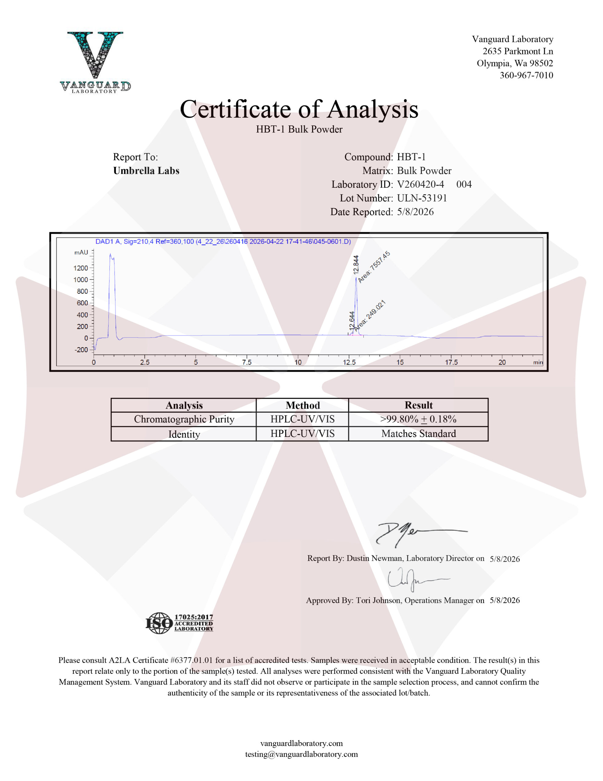
| File Name | View/Download |
| 2026-05-08-Umbrella-Labs-HBT-1-Certificate-of-Analysis-COA.pdf |  |
VIEW CERTIFICATES OF ANALYSIS (COA)
Additional information
| Weight | 1 oz |
|---|---|
| Dimensions | 1.5 × 1.5 × 1.5 in |
| Options |