PRE-084 HCL POWDER (1 GRAM)
$600.00
PRE-084 HCl is sold for laboratory research use only. Terms of sale apply. Not for human consumption, nor medical, veterinary, or household uses. Please familiarize yourself with our Terms & Conditions prior to ordering.

- Description
- Additional information
Description
PRE-084 HCl Nootropic Powder


| CAS Number | 75136-54-8 |
| Other Names | PRE-084 Hydrochloride, PRE 084 Hydrochloride, PRE084 Hydrochloride |
| IUPAC Name |
1-phenyl-2-(4-morpholinyl)ethyl ester-cyclohexanecarboxylic acid, monohydrochloride
|
| Molecular Formula | C₁₉H₂₈ClNO₃ |
| Molecular Weight | 353.88 |
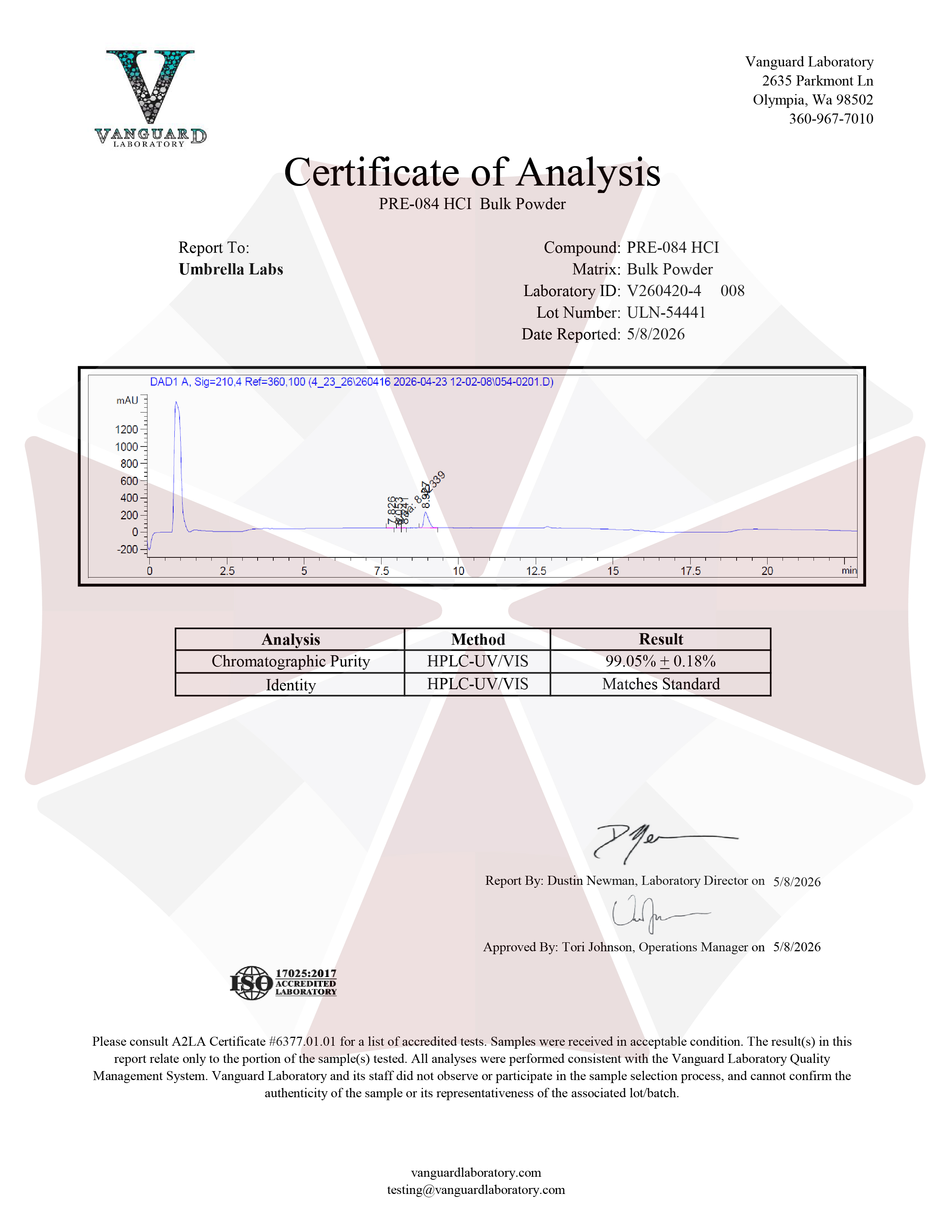
| Purity | ≥99% Pure (LC-MS) |
| Powder Availability |  1 gram |
| Storage | Store in cool dry environment, away from direct sunlight. |
| Terms | All products are for laboratory developmental research USE ONLY. Products are not for human consumption. |
What is PRE-084 HCl?
PRE-084 is a synthetic compound that has gained attention for its potential nootropic and neuroprotective properties due to its ability to selectively activate the sigma-1 receptor (S1R), a protein involved in regulating cellular stress responses, neurotransmission, and neuronal survival. The sigma-1 receptor plays an important role in maintaining proper communication between neurons, supporting calcium signaling, and protecting cells from oxidative damage and inflammation. Research suggests that activation of this receptor by PRE-084 may enhance cognitive processes by promoting synaptic plasticity, improving memory-related pathways, and supporting overall brain health. Preclinical studies have explored its potential benefits in models of neurodegenerative diseases, cognitive impairment, and neurological injury with results showing promising effects on learning, memory, and neuronal protection.
Main Research Findings
1) Activation of sigma-1 receptors by PRE-084 was found to reduce sepsis-related encephalopathy by targeting microglial inflammation and neuronal endoplasmic reticulum stress.
2) Stimulation of sigma-1 receptors via PRE-084 was shown to attenuate myocardial ischemia-reperfusion injury in an animal model.
Selected Data
1) The study performed by the research team of Zeng et al investigated the therapeutic potential of PRE-084, a Sigma-1 receptor (Sigma-1R) agonist, in reducing neurological damage associated with sepsis-associated encephalopathy (SAE). The researchers designed a controlled experimental study using a cecal ligation and puncture (CLP)-induced mouse model of sepsis to examine whether activation of Sigma-1R could protect against neuroinflammation, endoplasmic reticulum stress (ERS), synaptic damage, and cognitive impairment. Adult male and female C57BL/6J mice, aged 8–10 weeks and weighing approximately 20–25 grams, were used for the experiments. The animals were maintained under controlled environmental conditions, including regulated temperature, humidity, and light/dark cycles, with unrestricted access to food and water. Before experimentation, all animals underwent a seven-day acclimation period [1].
To establish sepsis-associated encephalopathy, researchers used the CLP surgical model, which is widely recognized for replicating the systemic inflammatory response observed during human sepsis. Mice were anesthetized using sodium pentobarbital before undergoing abdominal surgery. During the CLP procedure, the cecum was exposed, ligated with sterile silk sutures, and punctured twice using a 21-gauge needle to allow controlled leakage of intestinal contents into the abdominal cavity, creating polymicrobial infection. The cecum was then repositioned, and the abdominal incision was closed. Animals received warmed sterile saline after surgery and were monitored during recovery. Sham-operated control mice underwent the same surgical exposure but without ligation or puncture of the cecum, allowing researchers to distinguish the specific effects of sepsis from surgical stress.
The experimental design consisted of two main phases. The first phase examined how CLP-induced sepsis affected neurological outcomes, survival, and Sigma-1R expression in the brain. Mice were divided into sham and CLP groups, and neurological function was assessed over three postoperative days using the SHIRPA behavioral evaluation system. Survival was monitored for fourteen days after surgery. Brain tissues, particularly the hippocampus and cortex, were collected on postoperative days one and three to analyze Sigma-1R expression through Western blotting and immunofluorescence staining. These experiments helped identify whether Sigma-1R expression changed in response to sepsis and which brain regions were primarily affected [1].
The second phase evaluated the therapeutic effects of PRE-084 treatment. Animals were randomly assigned into four experimental groups: Sham plus saline, Sham plus PRE-084, CLP plus saline, and CLP plus PRE-084. Mice receiving treatment were administered PRE-084 at a dose of 10 mg/kg through intravenous tail vein injection immediately following surgery and again on postoperative days one and two. Control animals received equivalent saline injections. Researchers selected this dose based on previous evidence showing neuroprotective benefits of PRE-084 in neurological injury models and preliminary optimization experiments. Neurological assessments, survival analysis, biochemical testing, histological analysis, and cognitive testing were then performed to evaluate treatment outcomes [1].
Neurological impairment was measured using modified SHIRPA scoring, which assessed physical appearance, activity levels, posture, reflexes, gait abnormalities, tremors, respiratory function, touch response, and grip strength. Higher scores represented greater neurological impairment. Spatial learning and memory were evaluated using the Morris Water Maze between postoperative days fourteen and seventeen. During this test, mice were trained to locate a hidden underwater platform using visual cues. Researchers recorded escape latency, time spent in the target quadrant, platform crossings, and swimming speed to determine whether cognitive performance was affected by sepsis or improved through PRE-084 treatment.
To investigate the biological mechanisms responsible for PRE-084’s effects, multiple molecular techniques were performed. Western blot analysis measured Sigma-1R levels, synaptic proteins including PSD95 and synaptophysin, ER stress markers such as BiP and phosphorylated eIF2α, glial activation markers, and p38 MAPK pathway proteins. Immunofluorescence staining was used to identify Sigma-1R localization and quantify neuronal, microglial, and astrocyte responses. Markers including NeuN, Iba-1, GFAP, and phosphorylated p38 MAPK helped determine which cell types were involved in the inflammatory process. Enzyme-linked immunosorbent assays (ELISA) measured inflammatory cytokines, specifically tumor necrosis factor-alpha (TNF-α) and interleukin-6 (IL-6), in hippocampal and cortical tissue. Statistical analyses were performed using appropriate tests, including t-tests, ANOVA, repeated-measures analysis, and Kaplan–Meier survival comparisons. Overall, these methods allowed the researchers to comprehensively evaluate whether Sigma-1R activation through PRE-084 could improve neurological outcomes by regulating neuronal stress responses and neuroinflammation in SAE [1].
2) The study conducted by researchers Gao et al investigated whether activation of the sigma-1 receptor (σ1R) using the selective agonist PRE-084 could protect cardiac tissue from myocardial ischemia/reperfusion (I/R) injury and explored the possible molecular mechanisms responsible for these protective effects. The researchers focused specifically on whether PRE-084 could reduce cardiomyocyte apoptosis and improve cardiac function through activation of survival-related signaling pathways, particularly the Akt/endothelial nitric oxide synthase (eNOS) pathway. To evaluate this hypothesis, the investigators designed an in vivo animal experiment using adult male Sprague-Dawley rats and an established myocardial ischemia/reperfusion injury model. A total of forty-two healthy rats weighing between 220 and 250 grams were obtained from the animal center at Wuhan University and were used throughout the study [2].
The rats were randomly assigned using computer-generated allocation into three experimental groups, with fourteen animals included in each group. The first group served as the sham-operated control group, where rats underwent the same surgical procedures as experimental animals but without coronary artery occlusion. This allowed researchers to determine whether observed changes were specifically caused by ischemia/reperfusion injury rather than surgery itself. The second group was the I/R group, where rats underwent temporary coronary artery blockage followed by restoration of blood flow to reproduce myocardial injury caused by ischemia and subsequent reperfusion. The third group was the PRE-084 treatment group, where animals underwent the same I/R procedure but received PRE-084 pretreatment before surgery to determine whether σ1R stimulation could reduce myocardial damage.
To administer the experimental treatment, rats in the PRE-084 group received an intraperitoneal injection of PRE-084 dissolved in normal saline at a dose of 1 mg/kg one hour before myocardial ischemia was induced. Control animals received saline injections. The myocardial ischemia/reperfusion model was created surgically by temporarily blocking the left anterior descending coronary artery, a major vessel responsible for supplying blood to the heart. After anesthesia and surgical preparation, the coronary artery was ligated to prevent blood flow, creating myocardial ischemia. The occlusion was maintained for thirty minutes, after which the ligature was released to allow reperfusion for twenty-four hours. Successful reperfusion was confirmed by the recovery of ST-segment elevation, indicating restoration of coronary blood flow. This model allowed researchers to reproduce the injury that occurs clinically when blood flow returns after myocardial infarction treatment [2].
Following reperfusion, cardiac function was evaluated using hemodynamic measurements. Twenty-four hours after reperfusion, researchers inserted a polyethylene catheter through the right carotid artery and advanced it into the left ventricle. The catheter was connected to a multichannel physiological monitoring system that measured important indicators of heart function. These included left ventricular systolic pressure (LVSP), left ventricular end-diastolic pressure (LVEDP), and the maximum positive and negative rates of left ventricular pressure change (LV ± dp/dt). These parameters provided information regarding the heart’s ability to contract, relax, and maintain effective pumping function after injury [2].
To determine whether PRE-084 reduced myocardial cell death, researchers performed apoptosis analysis using terminal deoxynucleotidyl transferase-mediated dUTP nick-end labeling (TUNEL) staining. Five rats from each group were selected for this analysis. Heart tissue sections were treated with the TUNEL reaction mixture, fixed, stained, and examined under a light microscope. The apoptotic index was calculated by determining the percentage of apoptotic myocardial cells relative to the total number of cells. This method allowed researchers to quantify the extent of programmed cell death caused by I/R injury and determine whether PRE-084 provided protection.
Western blot analysis was performed to investigate the molecular pathways involved in PRE-084’s cardioprotective effects. Left ventricular tissue samples from five randomly selected animals in each group were homogenized, and proteins were extracted and quantified. Equal amounts of protein were separated using sodium dodecyl sulfate-polyacrylamide gel electrophoresis and transferred to membranes for antibody detection. Researchers measured proteins associated with apoptosis regulation, including Bax, Bcl-2, and cleaved caspase-3, as well as signaling proteins involved in cell survival, including p-Akt and phosphorylated eNOS (p-eNOS). Protein levels were quantified using imaging analysis and compared between groups. Statistical analysis was performed using SPSS software, with data reported as means and standard deviations. One-way analysis of variance followed by Student-Newman-Keuls testing was used to identify significant differences between groups. Overall, these methods allowed researchers to evaluate both functional and molecular evidence supporting the protective effects of PRE-084 against myocardial ischemia/reperfusion injury [2].
Discussion
1) The results of the study performed by Zeng et al demonstrated that PRE-084 treatment provided significant protection against sepsis-associated encephalopathy by improving survival, preserving neurological function, reducing neuronal stress, and suppressing neuroinflammation. Initial experiments confirmed that the CLP model successfully produced severe sepsis-related neurological impairment. Compared with sham-operated mice, CLP animals displayed significantly higher SHIRPA neurological deficit scores during the first three days following surgery, indicating reduced activity, abnormal behaviors, and impaired neurological responses. Survival analysis showed that CLP surgery dramatically reduced survival, with septic mice demonstrating a survival rate of approximately 33.3% over the fourteen-day monitoring period compared with 100% survival in sham animals. These findings confirmed the establishment of a clinically relevant SAE model [1].
The researchers next examined Sigma-1R expression following sepsis induction. Western blot analysis revealed that Sigma-1R protein levels significantly increased in the hippocampus three days after CLP surgery, while no significant differences were observed on the first postoperative day. Interestingly, Sigma-1R expression in the cortex remained unchanged, suggesting that the hippocampus may represent a particularly important brain region involved in Sigma-1R-mediated responses during SAE. Immunofluorescence staining further demonstrated that Sigma-1R was primarily located within neurons, showing strong overlap with the neuronal marker NeuN. In contrast, Sigma-1R showed minimal association with microglial marker Iba-1 or astrocyte marker GFAP, indicating that neurons were the primary site of Sigma-1R activity in this model.
Administration of PRE-084 significantly improved outcomes in septic mice. Although PRE-084 treatment did not further increase Sigma-1R expression levels, activation of the receptor produced substantial functional improvements. CLP mice treated with PRE-084 demonstrated improved neurological scores compared with untreated septic mice, particularly on postoperative days two and three. Survival rates also improved, increasing from 37.5% in saline-treated CLP mice to 62.5% in PRE-084-treated CLP mice. These findings suggested that activating existing Sigma-1R pathways, rather than increasing receptor expression itself, was responsible for the observed therapeutic effects [1].
Further analysis showed that PRE-084 protected against sepsis-induced synaptic damage. CLP surgery significantly decreased hippocampal expression of PSD95, a postsynaptic protein essential for maintaining synaptic structure and cognitive function. Treatment with PRE-084 significantly restored PSD95 levels, suggesting improved preservation of synaptic integrity. However, levels of synaptophysin, a presynaptic marker, were not significantly altered among groups. These results indicated that PRE-084 primarily protected postsynaptic structures that are vulnerable during septic brain injury [1].
The study also demonstrated that PRE-084 reduced endoplasmic reticulum stress, a major contributor to neuronal injury. Septic mice showed elevated levels of BiP and increased phosphorylation of eIF2α, indicating activation of harmful cellular stress pathways. PRE-084 administration significantly reduced both BiP expression and eIF2α phosphorylation in the hippocampus. Immunofluorescence analysis confirmed these findings by showing fewer p-eIF2α-positive cells in the hippocampal CA1 region and cortex after PRE-084 treatment. These results suggested that Sigma-1R activation protected neurons by reducing excessive ER stress responses caused by sepsis.
Cognitive testing using the Morris Water Maze revealed that PRE-084 improved sepsis-induced memory impairment. CLP mice treated with saline demonstrated longer escape latencies, reduced time spent in the target quadrant, and fewer platform crossings compared with sham mice, indicating impaired spatial learning and memory. In contrast, PRE-084-treated septic mice showed improved learning performance, spent significantly more time searching the correct target area, and crossed the former platform location more frequently. Swimming speeds were similar among groups, confirming that improved maze performance reflected better cognition rather than differences in physical ability [1].
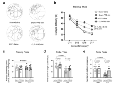
Figure 1: Changes in performance during the Morris water maze test in animals treated with saline vs treated with PRE-084.
Finally, PRE-084 significantly reduced neuroinflammatory responses associated with SAE. CLP surgery caused activation of microglia and astrocytes, demonstrated by increased Iba-1 and GFAP expression. PRE-084 treatment reduced both microglial and astrocytic activation. ELISA results showed that septic mice had elevated levels of inflammatory cytokines TNF-α and IL-6 in the hippocampus and cortex, while PRE-084 significantly lowered these inflammatory markers. Mechanistic analysis revealed that PRE-084 inhibited activation of the p38 MAPK inflammatory pathway, particularly within microglia. Overall, the findings demonstrated that PRE-084 protects against SAE by activating neuronal Sigma-1R, reducing ER stress, preserving synaptic proteins, suppressing microglial-driven inflammation, and ultimately improving survival and cognitive function [1].
2) The results reported by the research team of Gap et al demonstrated that stimulation of the sigma-1 receptor with PRE-084 significantly reduced myocardial ischemia/reperfusion injury by improving cardiac function, decreasing cardiomyocyte apoptosis, and activating protective molecular pathways. Initial evaluation focused on the ability of PRE-084 to preserve cardiac performance following ischemic injury. Hemodynamic measurements showed that ischemia/reperfusion caused substantial impairment in heart function compared with sham-operated animals. Rats exposed to I/R injury demonstrated reduced left ventricular contractile and relaxation capacity, indicating that temporary coronary artery occlusion followed by reperfusion produced significant myocardial dysfunction. However, rats treated with PRE-084 before ischemia showed meaningful improvements in cardiac performance. Compared with untreated I/R animals, PRE-084-treated rats exhibited significantly increased maximum positive and negative rates of left ventricular pressure change (LV ± dp/dt), reflecting improved myocardial contraction and relaxation ability. LVSP, an important indicator of the heart’s pumping strength, was also significantly improved after PRE-084 administration. LVEDP did not significantly differ between groups, suggesting that PRE-084’s primary benefits were related to improved contractile function rather than major changes in ventricular filling pressure [2].

Figure 2: Changes in hemodynamic and left ventricular functioning following treatment with PRE-084.
Because cardiomyocyte apoptosis is a major contributor to tissue damage following ischemia/reperfusion injury, the researchers next examined whether PRE-084 influenced programmed cell death. TUNEL staining revealed a significant increase in apoptotic myocardial cells in the I/R group compared with sham controls, confirming that ischemia followed by reperfusion triggered extensive cellular injury. In contrast, PRE-084 pretreatment significantly decreased the percentage of apoptotic cells compared with untreated I/R animals. These findings indicated that σ1R activation protected cardiac tissue by reducing the loss of cardiomyocytes after reperfusion injury. Since heart muscle cells have very limited regenerative ability, preventing apoptosis represents an important mechanism for preserving cardiac function following ischemic events.
To better understand the molecular mechanisms responsible for this reduction in apoptosis, the researchers evaluated proteins involved in regulating cell survival and cell death. Western blot analysis demonstrated that I/R injury disrupted the balance between pro-survival and pro-apoptotic proteins. Specifically, untreated I/R rats showed decreased expression of Bcl-2, an anti-apoptotic protein that protects cells from programmed death, and increased expression of Bax, a protein that promotes apoptosis. As a result, the protective Bcl-2/Bax ratio was significantly reduced after ischemia/reperfusion injury. Treatment with PRE-084 reversed these changes by increasing Bcl-2 expression while decreasing Bax expression, restoring a more favorable balance toward cell survival. These results suggested that PRE-084 reduced myocardial apoptosis by regulating key proteins controlling mitochondrial apoptotic pathways [2].
Additional analysis of apoptosis-related proteins revealed that PRE-084 influenced caspase-3 activation. Cleaved caspase-3, an important execution molecule involved in the final stages of apoptosis, was significantly elevated in the I/R group compared with sham animals. Increased cleaved caspase-3 indicated that ischemia/reperfusion activated cellular pathways leading to cardiomyocyte destruction. PRE-084 treatment significantly reduced cleaved caspase-3 levels, further confirming its ability to suppress apoptosis. Together, the TUNEL staining and Western blot findings provided strong evidence that σ1R stimulation reduced myocardial injury by limiting activation of programmed cell death pathways.
The researchers then examined whether activation of the Akt/eNOS signaling pathway contributed to PRE-084’s protective effects. Akt and eNOS are important molecules involved in promoting cell survival, regulating nitric oxide production, and protecting the heart from ischemic damage. Western blot results showed that myocardial ischemia/reperfusion significantly decreased phosphorylation of Akt and eNOS compared with sham controls, indicating suppression of this protective pathway after injury. However, PRE-084 treatment significantly increased expression of phosphorylated Akt and phosphorylated eNOS compared with untreated I/R rats. These findings suggested that PRE-084 activated the Akt/eNOS signaling cascade, which may contribute to improved cell survival and reduced myocardial damage [2].
The study concluded that PRE-084 provided significant cardioprotective effects during myocardial ischemia/reperfusion injury. By activating sigma-1 receptors, PRE-084 enhanced Akt/eNOS pathway signaling, increased survival-promoting proteins, decreased apoptosis-promoting molecules, and preserved cardiac function. The authors suggested that PRE-084 may represent a potential therapeutic candidate for reducing myocardial injury associated with ischemic heart diseases. However, they acknowledged limitations, including that PRE-084 was administered before ischemic injury rather than after myocardial infarction, which differs from most clinical situations. Additional research is required to determine whether PRE-084 remains effective when administered after ischemic events and to explore other possible mechanisms involving sigma-1 receptor interactions with calcium, potassium, sodium, and chloride channels. Overall, the findings provided strong experimental evidence that PRE-084 protects the heart primarily through anti-apoptotic effects mediated by activation of Akt/eNOS signaling pathways [2].
Disclaimer
**LAB USE ONLY**
*This information is for educational purposes only and does not constitute medical advice. THE PRODUCTS DESCRIBED HEREIN ARE FOR RESEARCH USE ONLY. All clinical research must be conducted with oversight from the appropriate Institutional Review Board (IRB). All preclinical research must be conducted with oversight from the appropriate Institutional Animal Care and Use Committee (IACUC) following the guidelines of the Animal Welfare Act (AWA).
Citations
[1] Zeng X, Kang W, Zhou Q, Pan X, Wang L. Sigma-1 receptor activation by PRE-084 attenuates sepsis-associated encephalopathy by targeting microglial p38 MAPK-mediated neuroinflammation and neuronal endoplasmic reticulum stress. Inflamm Res. 2025;74(1):117. Published 2025 Sep 4. doi:10.1007/s00011-025-02086-5
[2] Gao QJ, Yang B, Chen J, Shi SB, Yang HJ, Liu X. Sigma-1 Receptor Stimulation with PRE-084 Ameliorates Myocardial Ischemia-Reperfusion Injury in Rats. Chin Med J (Engl). 2018;131(5):539-543. doi:10.4103/0366-6999.226076
PRE-084 HCl is sold for laboratory research use only. Terms of sale apply. Not for human consumption, nor medical, veterinary, or household uses. Please familiarize yourself with our Terms & Conditions prior to ordering.


 The Potential Of PRE-084: A Profile Of Possibilities


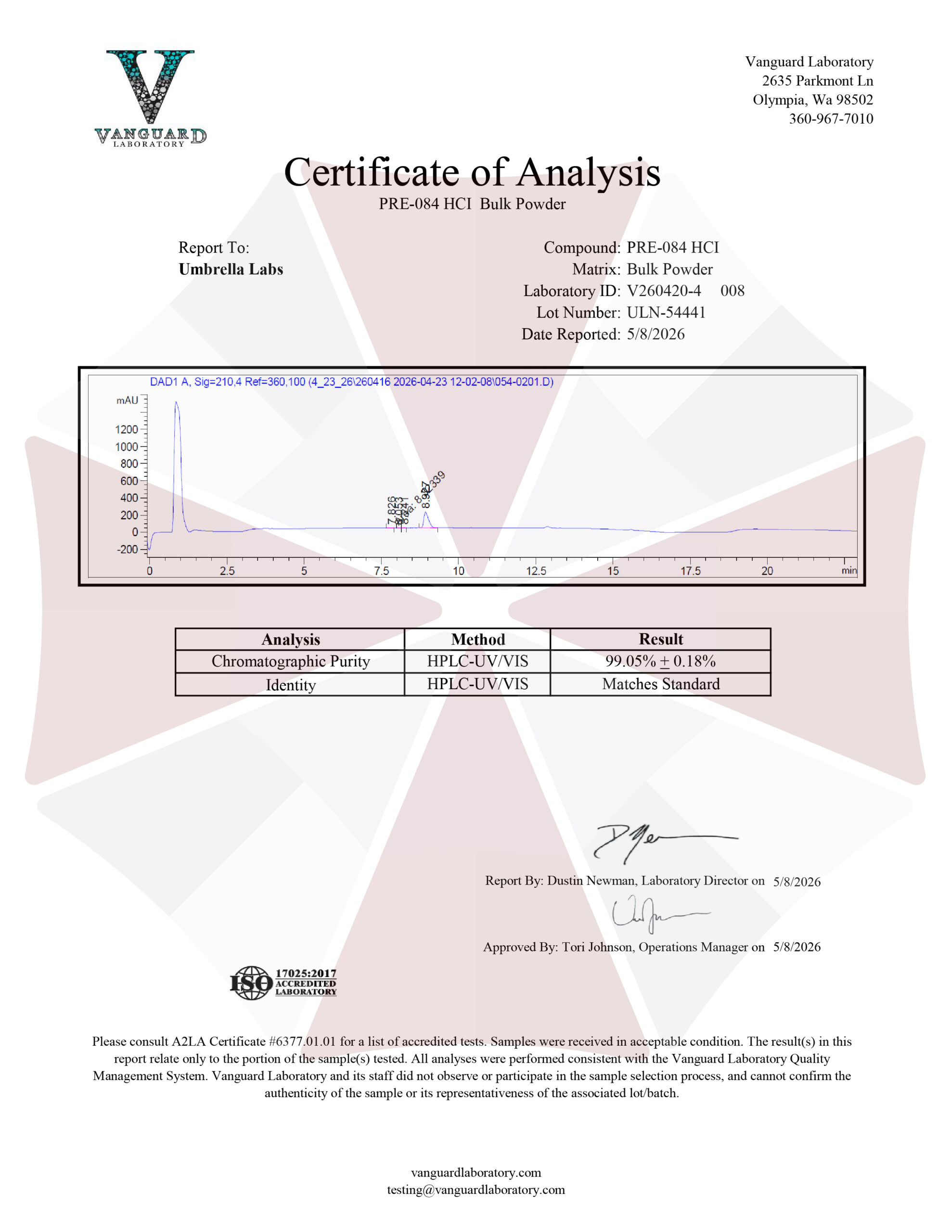
| File Name | View/Download |
| 2026-05-08-Umbrella-Labs-PRE-084-HCl-Certificate-of-Analysis-COA.pdf | |
VIEW CERTIFICATES OF ANALYSIS (COA)
Additional information
| Weight | 1 oz |
|---|---|
| Dimensions | 1.5 × 1.5 × 1.5 in |
| Weight | 1 Gram, 5 Grams, 10 Grams |