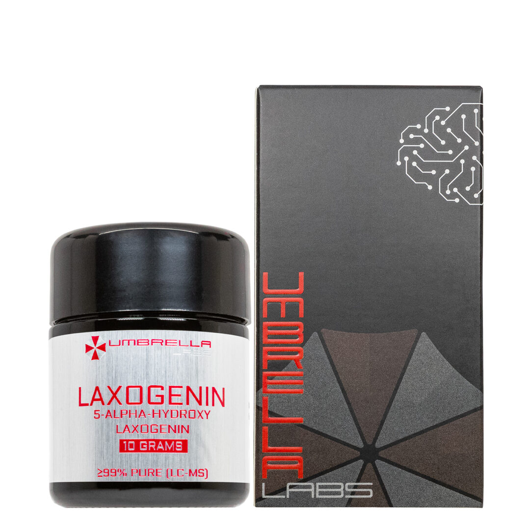
S107 POWDER (1 GRAM)
$199.99
S107 is sold for laboratory research use only. Terms of sale apply. Not for human consumption, nor medical, veterinary, or household uses. Please familiarize yourself with our Terms & Conditions prior to ordering.

- Description
- Additional information
Description
S107 Nootropic Powder


| CAS Number | 927871-76-9 |
| Other Names | S107, S-107, S 107 |
| IUPAC Name | 2,3,4,5-Tetrahydro-7-methoxy-4-methyl-1,4-benzothiazepine hydrochloride |
| Molecular Formula | C₁₁H₁₅NOS |
| Molecular Weight | 209.31 |
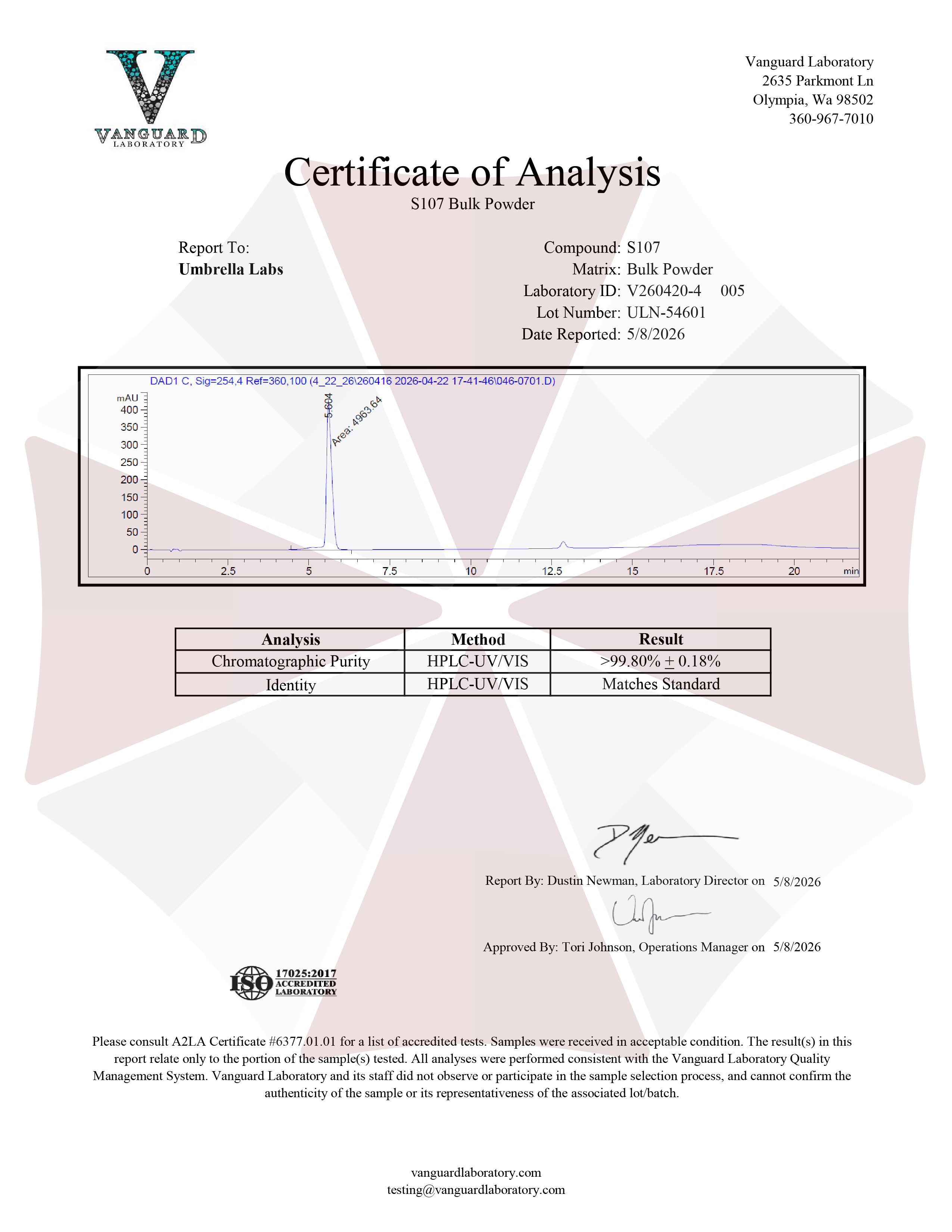
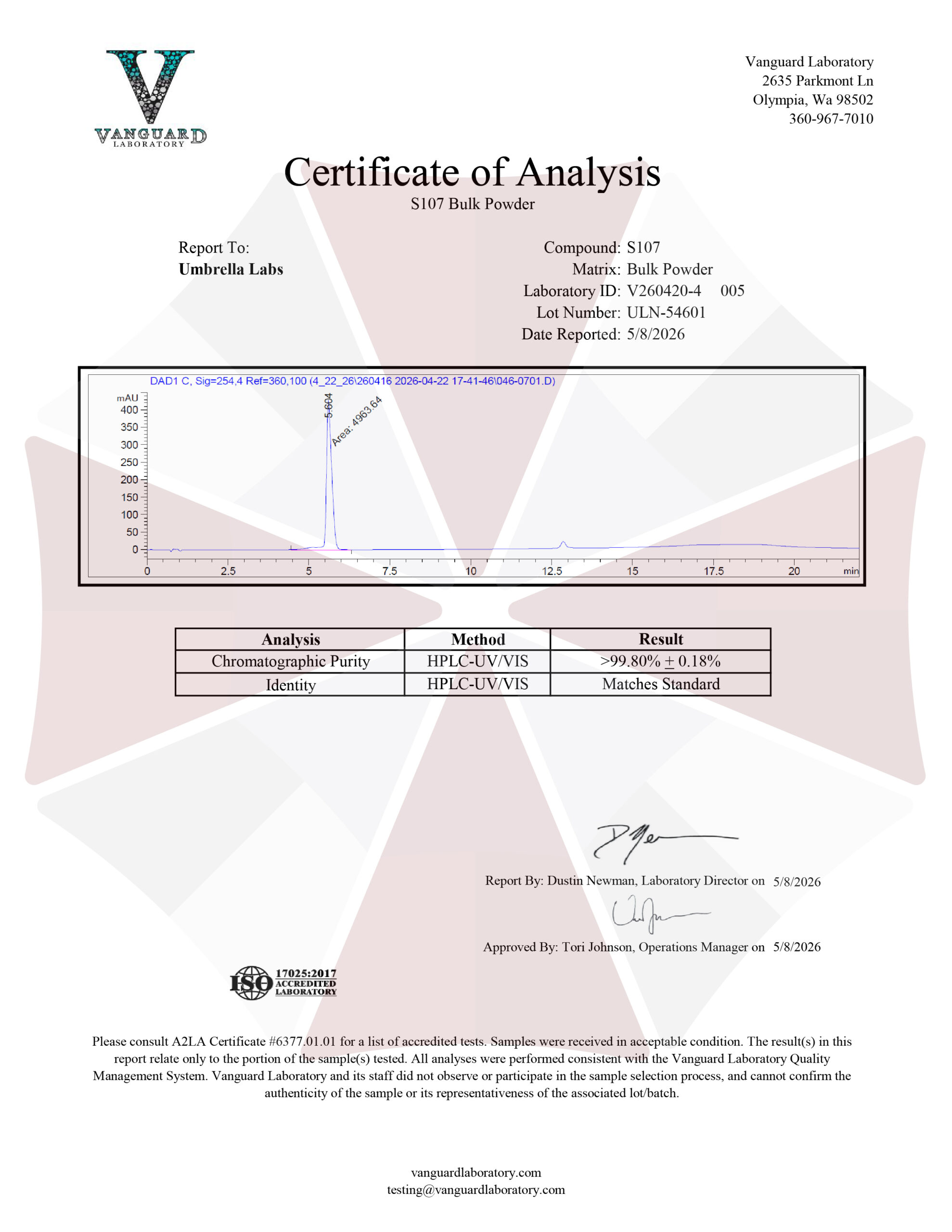
| Purity | ≥99% Pure (LC-MS) |
| Powder Availability |  1 gram |
| Storage | Store in cool dry environment, away from direct sunlight. |
| Terms | All products are for laboratory developmental research USE ONLY. Products are not for human consumption. |
What is S107?
S107 is a small-molecule ryanodine receptor (RyR) stabilizer that has attracted interest as a potential nootropic and neuroprotective compound because of its ability to regulate intracellular calcium signaling. Rather than acting directly on conventional neurotransmitter receptors, S107 is designed to stabilize the interaction between ryanodine receptors and their associated calcium-channel regulatory protein, calstabin, thereby reducing abnormal calcium leakage from intracellular stores. Dysregulated calcium signaling and excessive intracellular calcium release have been implicated in neuronal dysfunction, oxidative stress, synaptic abnormalities, and impaired cellular function. By promoting more stable RyR channel activity, S107 has been investigated as a means of preserving normal calcium homeostasis and protecting neuronal and synaptic function. Preclinical research has demonstrated particular interest in S107 in models of neurological and neurodegenerative disorders, where abnormal RyR activity may contribute to synaptic dysfunction. In experimental studies involving amyloid-beta-induced GABAergic abnormalities, S107 counteracted increases in inhibitory synaptic transmission and restored abnormalities in synchronous and asynchronous neurotransmitter release, supporting the possibility that RyR stabilization may have therapeutic relevance for maintaining neuronal signaling.
Main Research Findings
1) GABAergic synaptic alterations related to Abeta42 oligomers were inhibited in hippocampal neurons following treatment with S107.
2) S107 was shown to improve contractility in an animal model of knockout cardiomyocytes.
Selected Data
1) The research team Hidisoglu et al. investigated the effects of amyloid beta 42 (Abeta42) oligomers on GABAergic synaptic transmission in hippocampal neurons and examined whether S107, a ryanodine receptor (RyR)–calstabin interaction stabilizer, could prevent or reverse the resulting synaptic abnormalities. The experimental design focused on determining whether Abeta42 altered GABAergic transmission through presynaptic mechanisms, postsynaptic mechanisms, or both. Particular attention was given to spontaneous miniature inhibitory postsynaptic currents (mIPSCs), evoked inhibitory postsynaptic currents (eIPSCs), short-term synaptic plasticity, neurotransmitter release probability, quantal properties, the readily releasable pool of synaptic vesicles, and the balance between synchronous and asynchronous GABA release. The researchers also evaluated whether stabilizing RyR function with S107 could restore these parameters toward control levels. The study was performed using primary hippocampal neurons derived from C57BL6 mice, allowing the investigators to examine the direct cellular effects of Abeta42 in a controlled experimental environment [1].
Hippocampal neurons were isolated from embryonic day 18 C57BL6 mouse embryos. Embryos were removed by caesarean section and rapidly decapitated, after which the hippocampi were dissected under sterile conditions. The tissue was maintained in cold, high-glucose Hanks’ balanced salt solution (HBSS) and enzymatically digested using papain at 0.5 mg/ml in HBSS together with DNase at 0.1 mg/ml. Following dissociation, the isolated hippocampal neurons were plated at a density of approximately 100 cells/mm². Cells were maintained in Neurobasal culture medium supplemented with B-27, glutamine, and penicillin-streptomycin. Half of the culture medium was replaced every seven days, and the neurons were maintained for approximately two to three weeks before electrophysiological experiments were performed.
The researchers then established pharmacological conditions designed to isolate GABAergic currents. The extracellular Tyrode’s solution contained CaCl₂, NaCl, MgCl₂, HEPES, KCl, and glucose, while the intracellular pipette solution contained CsCl, TEACl, EGTA, glucose, MgCl₂, ATP, GTP, and phosphocreatine. Several receptor antagonists were incorporated into the extracellular solution to prevent contamination from other neurotransmitter systems. d-AP5 was used to block NMDA receptors, DNQX was used to inhibit non-NMDA glutamate receptors, and CGP58845 was used to block GABAB receptors. Tetrodotoxin (TTX) was added during mIPSC experiments to inhibit spontaneous action-potential propagation. For evoked recordings, QX-314 was also included in the intracellular solution to block postsynaptic sodium currents. Abeta42 was prepared as a 1 mM stock solution in 1% ammonium hydroxide, and cultured neurons were exposed to 1 μM Abeta42 for 48 hours before electrophysiological recordings. For experiments investigating RyR stabilization, neurons were treated with 10 μM S107 for 48 hours together with Abeta42 [1].
Whole-cell patch-clamp recordings were performed using borosilicate glass electrodes with a resistance of 3–5 MΩ. Neurons were held at −70 mV. Electrophysiological signals were sampled at 10 kHz and filtered using an eight-pole low-pass Bessel filter. Recordings were excluded when leak current exceeded 100 pA or series resistance was greater than 20 MΩ. mIPSCs were analyzed using Mini Analysis software, with a 10-pA amplitude threshold for event detection. The researchers measured both mIPSC amplitude and frequency and performed variance analysis to estimate the unitary current generated by individual GABAA receptors and the number of receptors activated by each quantum of GABA.
Evoked monosynaptic GABAergic responses were recorded from pairs of hippocampal neurons maintained for 14–21 days in vitro. Presynaptic stimulation was delivered through a glass pipette positioned against the soma of a GABAergic interneuron. Brief electrical pulses were used to produce eIPSCs, which were recorded at regular intervals. The researchers examined eIPSC amplitude, total charge, and deactivation kinetics. To investigate presynaptic function, paired-pulse depression was measured using two consecutive stimuli separated by interpulse intervals ranging from 25 ms to 2 seconds. Multiprobability fluctuation analysis was subsequently performed by altering extracellular Cd²⁺ concentrations. This approach allowed the investigators to estimate the number of independent release sites, average quantal size, and average probability of vesicle release [1].
The researchers also examined vesicle-pool dynamics using high-frequency stimulation. Neurons received 80 repetitive stimuli at 40 Hz for two seconds, allowing analysis of the synchronous readily releasable pool (RRPsyn), vesicle release probability, and asynchronous release. Delayed asynchronous release was measured by quantifying the current occurring during the one-second period following the stimulation train. The total readily releasable pool (RRPtot), which supplies vesicles for both synchronous and asynchronous release, was estimated from cumulative eIPSC charge. Statistical analyses included Student’s t-tests and one-way ANOVA with Tukey’s test for normally distributed data, while Mann–Whitney U, Kolmogorov–Smirnov, and Kruskal–Wallis tests with Dunn’s post hoc analysis were used for non-normally distributed data [1].
2) The research team of Guo et al. investigated the progression of cardiac dysfunction caused by loss of RNA binding motif 20 (RBM20) and examined whether the ryanodine receptor stabilizer S107 could improve abnormal calcium handling and cardiomyocyte contractility. RBM20 is a cardiac-enriched splicing factor that regulates the alternative splicing of titin and several proteins involved in calcium handling. The investigators used wild-type (WT) and homozygous Rbm20 knockout (KO) rats to determine how RBM20 depletion affected cardiac structure, myocardial stiffness, intracellular calcium regulation, and cardiomyocyte mechanical function across different ages. They evaluated rats at 3, 6, 9, and 12 months to establish when the overt dilated cardiomyopathy (DCM) phenotype developed. They also conducted both in vitro and in vivo S107 treatment experiments to determine whether stabilizing the RyR2–calstabin complex could reduce abnormal calcium release and improve cardiac performance [2].
The experimental animals consisted of male WT and Rbm20 KO rats between 3 and 12 months of age. The KO animals were derived from a spontaneous mutant and were generated from Sprague-Dawley × Brown Norway rats. Animals were maintained on standard rodent chow, and the experimental procedures were performed under institutional animal-care approval. For the in vivo S107 experiment, 12-month-old male WT rats received normal drinking water, while KO rats received drinking water containing S107 at 20 mg/kg/day. The KO animals were treated for two weeks. Echocardiography was performed before treatment and again after the 14-day treatment period. This design allowed the researchers to determine whether S107 could modify established cardiac abnormalities in older KO animals.
Cardiac structure and function were assessed using two complementary approaches. Echocardiography was performed in WT and KO rats at 3, 6, 9, and 12 months using two-dimensional guided M-mode imaging and a 15–6 MHz linear transducer. Measurements were used to evaluate ventricular dimensions, wall thickness, and fractional shortening. In addition, pressure-volume loop analysis was performed to provide a more detailed assessment of ventricular performance. Rats were anesthetized with ketamine and xylazine, and vascular catheters were placed in the femoral and jugular veins. A pressure-volume microcatheter was introduced through the carotid artery into the left ventricle. Following a stabilization period, transient inferior vena cava occlusion was used to generate pressure-volume loops over a range of end-diastolic volumes. Hypertonic saline was administered to permit parallel-conductance correction, and the resulting loops were analyzed using specialized acquisition and PV-loop software [2].
To examine myocardial mechanical properties directly, the investigators isolated myofibrils from the left ventricles of 3-month-old WT and KO rats. Ventricular tissue was chemically skinned using Triton-X in rigor solution containing protease inhibitors before being homogenized in relaxing solution. Individual bundles of approximately 7–10 myofibrils were mounted between a motor-controlled micro-tool and a calibrated force probe. The myofibrils were progressively stretched, and resting tension was recorded at different sarcomere lengths. Sarcomere length and myofibril diameter were measured using ImageJ, while the mechanical data were collected and analyzed using customized LabView software. Measurements from myofibrils belonging to the same animal were averaged within 0.15-μm sarcomere-length intervals and subsequently used to characterize the relationship between sarcomere length and resting tension [2].
Adult cardiomyocytes were isolated from approximately 6-month-old rats to investigate cellular contractility and calcium dynamics. Hearts were removed under deep anesthesia and cannulated through the aorta. They were initially perfused with oxygenated physiological buffer and subsequently with a digestion solution containing collagenase II, trypsin, and calcium. Once the heart became sufficiently flaccid, digestion was stopped, the left ventricle was dissected, and the tissue was mechanically triturated and filtered. Calcium was gradually reintroduced until the cardiomyocytes were prepared for experiments. Rod-shaped cardiomyocytes with clear edges were selected for contractility measurements. Cells were incubated with 10 μM freshly prepared S107 for 2–3 hours at room temperature, while vehicle-treated cells served as controls. The cells were placed in an IonOptix SoftEdge Myocam system and electrically stimulated at 1 Hz with 3-ms pulses at 15 V. Peak shortening, shortening duration, time to 50% relengthening, and maximal velocities of shortening and relengthening were recorded.
Intracellular calcium dynamics were assessed using Fura-2 AM fluorescence. Cardiomyocytes were loaded with 0.5 μM Fura-2 AM for 10 minutes and analyzed using dual-excitation fluorescence at 360 and 380 nm. Cells were electrically stimulated at 0.5 Hz, and fluorescence was sampled at 333 Hz. The ratio of fluorescence intensity at the two excitation wavelengths was used to assess intracellular calcium levels, while the decay time was analyzed as an indicator of calcium clearance. The researchers also characterized molecular changes associated with RBM20 depletion. Titin isoforms from 3-, 6-, and 9-month-old hearts were separated using SDS-agarose gel electrophoresis. Western blotting of 6-month-old heart tissue measured total RyR2 and phosphorylated RyR2 at Ser2808, while RT-PCR examined Ryr2 transcript variants involving exons 76–77 [2].
Discussion
1) The results from the study performed by Hidisoglu et al demonstrated that Abeta42 oligomers substantially altered GABAergic synaptic transmission in cultured hippocampal neurons. The first experiments examined spontaneous miniature inhibitory postsynaptic currents, which provide information about individual synaptic events and can reveal changes in both presynaptic neurotransmitter release and postsynaptic receptor function. Abeta42 significantly shifted the cumulative distribution of mIPSC amplitude toward larger values and shifted the distribution of inter-event intervals toward shorter intervals. Quantitatively, mean mIPSC amplitude increased from 23.74 ± 6.09 pA in control neurons to 32.78 ± 8.41 pA following Abeta42 exposure. At the same time, the mean inter-event interval decreased from 988.3 ± 412.9 ms to 391.5 ± 86.9 ms. These changes demonstrated that Abeta42 increased both the strength and frequency of spontaneous GABAergic events. The decay slope of the mIPSCs was not altered, indicating that Abeta42 did not produce a major change in the basic deactivation kinetics of GABAA receptors [1].
The researchers then used variance analysis to investigate the postsynaptic mechanisms underlying the increased mIPSC amplitude. Interestingly, Abeta42 reduced the estimated unitary current of individual GABAA receptor channels. The unitary current decreased from 4.28 ± 2.14 pA in control neurons to 2.93 ± 1.22 pA following Abeta42 treatment. Despite this decrease, the estimated number of GABAA receptor channels activated by a single quantum of neurotransmitter increased substantially, from 6.98 ± 3.78 in control neurons to 13.83 ± 7.90 in Abeta42-treated neurons. Therefore, the larger mIPSCs were not caused by individual GABAA channels producing stronger currents. Instead, the increase was associated with activation of a greater number of receptors by each quantum of GABA. Combined with the increased frequency of mIPSCs, these findings suggested that Abeta42 affects both postsynaptic receptor organization and presynaptic processes involved in spontaneous neurotransmitter release.
Evoked GABAergic transmission was also significantly affected by Abeta42. The average amplitude of eIPSCs increased from 732.71 ± 370.40 pA in control neurons to 1280.04 ± 375.90 pA after Abeta42 exposure. The area or charge underlying the eIPSC similarly increased from 58.55 ± 32.27 pC to 137.31 ± 71.73 pC. However, the fast and slow components of eIPSC deactivation were not significantly different between control and Abeta42-treated neurons. This indicated that the increased eIPSC response was primarily related to an increase in synaptic response magnitude rather than slower GABAA receptor deactivation. Importantly, S107 substantially counteracted the Abeta42-induced increase. In neurons treated with both Abeta42 and S107, eIPSC amplitude decreased to 616.60 ± 313 pA and eIPSC charge decreased to 60.52 ± 47.34 pC, values comparable to control neurons. S107 did not significantly alter the deactivation time constants [1].
To determine whether the increased eIPSC amplitude resulted from greater presynaptic release probability, the investigators examined paired-pulse depression. Control synapses exhibited approximately 70.55% depression when the interpulse interval was 25 ms. Abeta42-treated neurons showed 68.98% depression at the same interval, which was not significantly different from control. The relationship between paired-pulse depression and longer interpulse intervals was also similar between groups. These results indicated that Abeta42 did not significantly alter short-term synaptic plasticity or the probability of neurotransmitter release. Multiprobability fluctuation analysis provided additional evidence that other presynaptic properties were responsible for the increased eIPSC amplitude. Abeta42 increased the estimated number of independent GABAergic release sites from 171.59 ± 103.1 to 264.70 ± 117.3 and increased average quantal size from 13.16 ± 4.61 to 17.72 ± 7.19 pA. In contrast, average release probability remained unchanged at approximately 0.66 in controls and 0.69 following Abeta42 treatment.
High-frequency stimulation revealed that Abeta42 altered the balance between synchronous and asynchronous GABA release. Delayed asynchronous release was significantly reduced following Abeta42 exposure. In control neurons, delayed asynchronous release was approximately 19.01 ± 11.44 times the synchronous release reference, whereas Abeta42-treated neurons showed a value of only 5.75 ± 4.22 times the corresponding reference. The normalized increase in asynchronous release during stimulation was also significantly reduced by Abeta42, with the slope changing from 0.124 ± 0.006 in controls to −0.0129 ± 0.0145 after treatment. S107 reversed this effect, increasing delayed asynchronous release to 19.66 ± 16.05 times the corresponding synchronous charge, which was comparable to control values [1].
Despite these changes, the total readily releasable pool was not significantly affected by Abeta42. RRPtot was 188.75 ± 177.01 pC in control neurons compared with 216.17 ± 140.39 pC after Abeta42 treatment. S107 also produced no significant effect on RRPtot. In contrast, the synchronous readily releasable pool was strongly increased. RRPsyn increased from 1897.01 ± 1652 pA in control neurons to 5193.02 ± 3961 pA following Abeta42 exposure. The probability of vesicle release from this pool remained unchanged, indicating that the larger synchronous response was associated with a larger pool of readily available vesicles rather than an increased probability that individual vesicles would be released. S107 reduced RRPsyn to 1813 ± 1532 pA, restoring it toward control levels [1].
Overall, the findings showed that Abeta42 oligomers produce a complex alteration of hippocampal GABAergic synapses involving both presynaptic and postsynaptic mechanisms. Abeta42 increased spontaneous GABAergic activity, increased GABAA receptor recruitment, increased the number of independent release sites, increased quantal size, and enlarged the pool of vesicles available for synchronous release. At the same time, it reduced delayed asynchronous GABA release without changing total vesicle-pool size or overall release probability. Treatment with S107 reversed many of these abnormalities, including the increased eIPSC amplitude, enlarged synchronous readily releasable pool, and reduced asynchronous release. The research team therefore concluded that RyR dysfunction contributes to Abeta42-induced GABAergic synaptic impairment and that stabilizing RyR–calstabin interactions with S107 may represent a potential strategy for preventing or treating early synaptic abnormalities associated with Alzheimer’s disease [1].
2) The study completed by Guo et al demonstrated that RBM20 depletion produced age-dependent alterations in titin expression, myocardial stiffness, cardiac morphology, calcium regulation, and cardiomyocyte contractility. The earliest changes were observed in titin isoform expression and passive mechanical properties, whereas the overt DCM phenotype developed later. Titin gel electrophoresis showed that WT hearts predominantly expressed the smaller and stiffer N2B titin isoform at 3, 6, and 9 months, whereas Rbm20 KO hearts expressed a larger N2BA-G isoform. This isoform shift was accompanied by reduced passive mechanical tension. When myofibrils were isolated from 3-month-old animals, those from KO hearts produced significantly lower resting tension than myofibrils from WT animals. Pressure-volume loop measurements similarly demonstrated significantly decreased diastolic stiffness in 3-month-old KO rats. Thus, RBM20 loss altered titin composition and myocardial mechanical properties before the development of obvious ventricular dysfunction [2].
The cardiac phenotype became progressively more severe with age. At three months, there were no significant differences between WT and KO rats in left ventricular end-diastolic internal dimension, posterior wall thickness, end-systolic internal dimension, end-systolic posterior wall thickness, or fractional shortening. However, substantial structural and functional abnormalities emerged at six months and persisted at nine and twelve months. KO rats developed enlarged ventricular chambers, thinner posterior walls, and reduced fractional shortening compared with WT animals. Pressure-volume loop analysis additionally demonstrated decreased cardiac output in KO rats at six and nine months. These findings indicated that loss of RBM20 initially altered myocardial stiffness but required additional time to produce the overt ventricular dilation and contractile dysfunction characteristic of DCM. The researchers therefore identified approximately six months of age as the point at which the KO rats began displaying a clearly established cardiomyopathic phenotype [2].
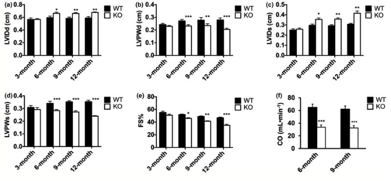
Figure 1: Changes in the parameters of cardiac function following treatment with S107.
Molecular analysis suggested that abnormal RyR2 regulation contributed to the cardiac phenotype. Western blotting demonstrated that total RyR2 protein levels were not significantly altered by RBM20 depletion, but phosphorylation of RyR2 at Ser2808 was significantly increased in KO hearts. RT-PCR also showed increased expression of a larger Ryr2 transcript variant in KO animals. Because increased RyR2 phosphorylation can destabilize the RyR2–FKBP12.6, or calstabin 2, complex, the researchers proposed that RBM20 loss promoted abnormal RyR2 activity and increased sarcoplasmic reticulum calcium leakage. These molecular changes provided a potential mechanistic connection between RBM20-dependent RNA splicing abnormalities and the impaired calcium regulation observed in KO cardiomyocytes.
The isolated cardiomyocyte experiments confirmed significant impairments in contraction. Rbm20 KO cardiomyocytes exhibited reduced peak shortening and reduced maximum velocity of sarcomere shortening compared with WT cells. The time to 50% peak shortening was also altered, indicating abnormal contraction kinetics. Importantly, treatment with 10 μM S107 restored peak shortening, shortening velocity, and the time to 50% peak shortening in KO cardiomyocytes to levels comparable with WT cells. In contrast, RBM20 depletion did not significantly alter maximal velocity of relengthening or the time to 50% relengthening, and S107 did not affect these parameters. These findings suggested that the primary mechanical deficit caused by RBM20 depletion involved contraction rather than relaxation. The research team associated the impaired shortening response with abnormal calcium release rather than defective calcium uptake [2].
Measurements of intracellular calcium provided further evidence for impaired RyR2-mediated calcium regulation. Rbm20 KO cardiomyocytes displayed significantly elevated baseline Fura-2 fluorescence, indicating increased resting intracellular calcium levels. At the same time, electrically stimulated increases in Fura-2 fluorescence were suppressed, demonstrating an abnormal calcium response during stimulation. Importantly, the rate of intracellular calcium decay did not differ between WT and KO cardiomyocytes, suggesting that calcium removal or uptake into the sarcoplasmic reticulum was not substantially impaired. S107 treatment eliminated the elevated baseline fluorescence in KO cardiomyocytes and improved the electrically stimulated calcium increase without significantly affecting calcium decay. These results supported the conclusion that RBM20 depletion primarily caused excessive resting calcium release through dysfunctional RyR2 channels. Stabilization of the RyR2 complex with S107 reduced this calcium leak and improved the ability of the cardiomyocytes to generate a normal calcium response.
Overall, the study showed that RBM20 loss produces a progressive cardiac phenotype involving early titin isoform switching and reduced myocardial stiffness followed by ventricular dilation and impaired cardiac function at older ages. S107 successfully corrected abnormal resting calcium levels and contractile deficits in isolated adult KO cardiomyocytes, supporting a role for RyR2-mediated calcium leakage in RBM20-associated dysfunction. However, the lack of improvement following in vivo treatment suggested that calcium dysregulation is only one component of RBM20 cardiomyopathy. The researchers noted that RBM20 regulates the splicing of numerous additional cardiac genes, meaning that abnormalities in titin, calcium handling, ion transport, and sarcomere structure may collectively contribute to disease progression. They also emphasized that treatment began at 12 months, well after the overt DCM phenotype emerged around six months, and that the two-week treatment period may have been insufficient. Consequently, the study supports S107 as a promising approach for correcting cellular calcium-handling abnormalities [2] .
Disclaimer
**LAB USE ONLY**
*This information is for educational purposes only and does not constitute medical advice. THE PRODUCTS DESCRIBED HEREIN ARE FOR RESEARCH USE ONLY. All clinical research must be conducted with oversight from the appropriate Institutional Review Board (IRB). All preclinical research must be conducted with oversight from the appropriate Institutional Animal Care and Use Committee (IACUC) following the guidelines of the Animal Welfare Act (AWA).
Citations
[1] Hidisoglu E, Chiantia G, Franchino C, et al. The ryanodine receptor-calstabin interaction stabilizer S107 protects hippocampal neurons from GABAergic synaptic alterations induced by Abeta42 oligomers. J Physiol. 2022;600(24):5295-5309. doi:10.1113/JP283537
[2] Guo W, Zhu C, Yin Z, et al. The ryanodine receptor stabilizer S107 ameliorates contractility of adult Rbm20 knockout rat cardiomyocytes. Physiol Rep. 2021;9(17):e15011. doi:10.14814/phy2.15011
S107 is sold for laboratory research use only. Terms of sale apply. Not for human consumption, nor medical, veterinary, or household uses. Please familiarize yourself with our Terms & Conditions prior to ordering.


| File Name | View/Download |
| 2026-05-08-Umbrella-Labs-S107-Certificate-of-Analysis-COA.pdf |  |
VIEW CERTIFICATES OF ANALYSIS (COA)
Additional information
| Weight | 1 oz |
|---|---|
| Dimensions | 1.5 × 1.5 × 1.5 in |
| Options |