


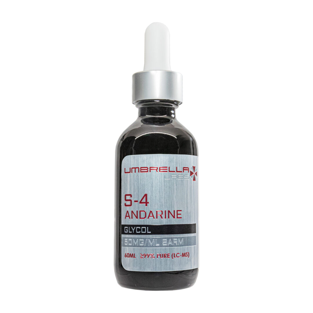


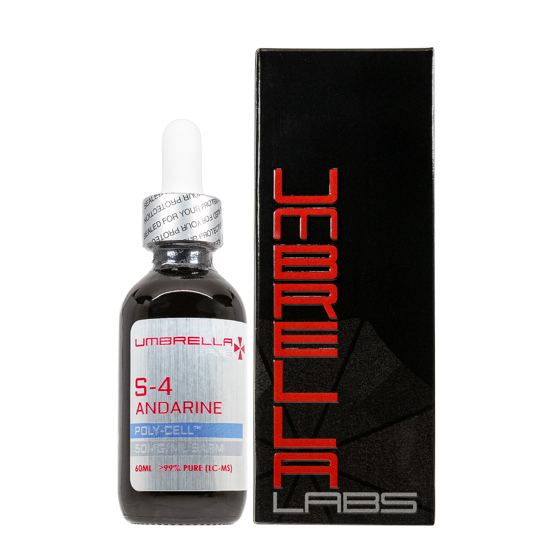

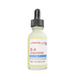
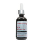
S-4 ANDARINE SARM – 50MG/ML – 30ML/60ML BOTTLE For Sale

$70.99 – $154.98
S-4 Andarine SARM is sold for laboratory research use only. Terms of sale apply. Not for human consumption, nor medical, veterinary, or household uses. Please familiarize yourself with our Terms & Conditions prior to ordering.

Also Available In:
- Description
- Additional information
Description
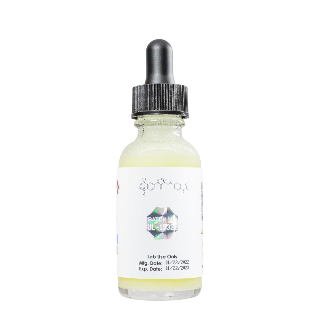
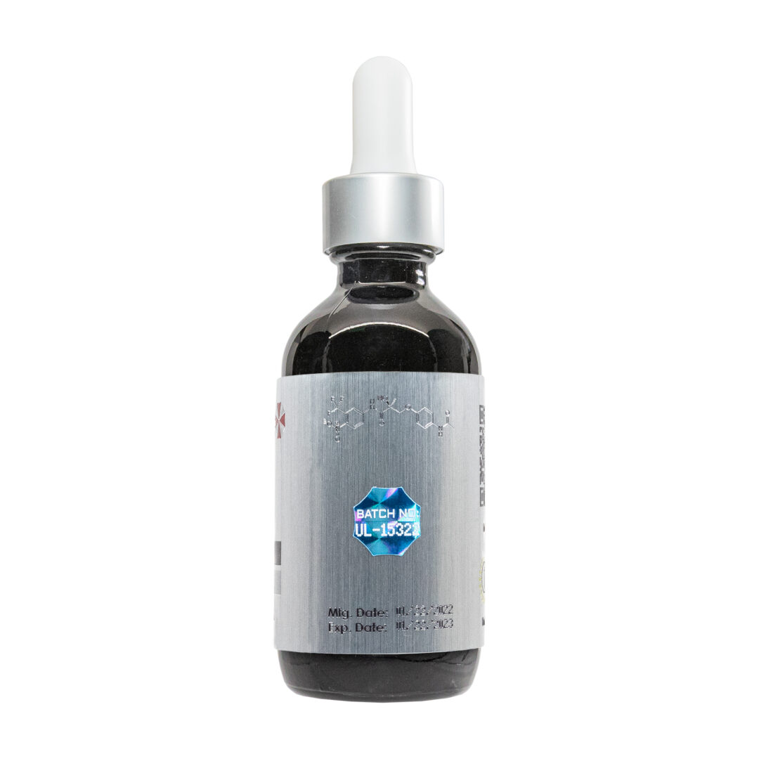
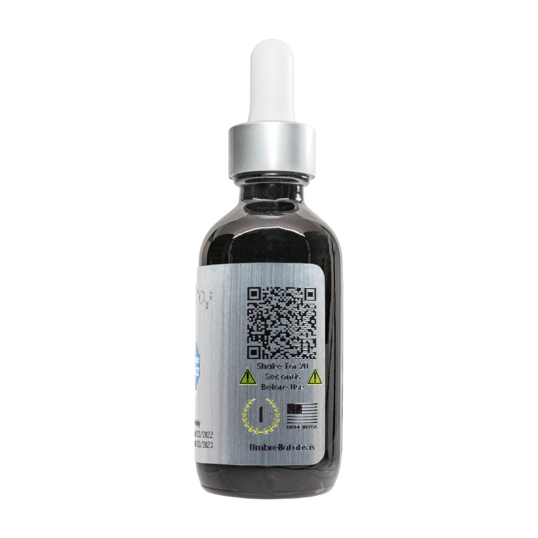
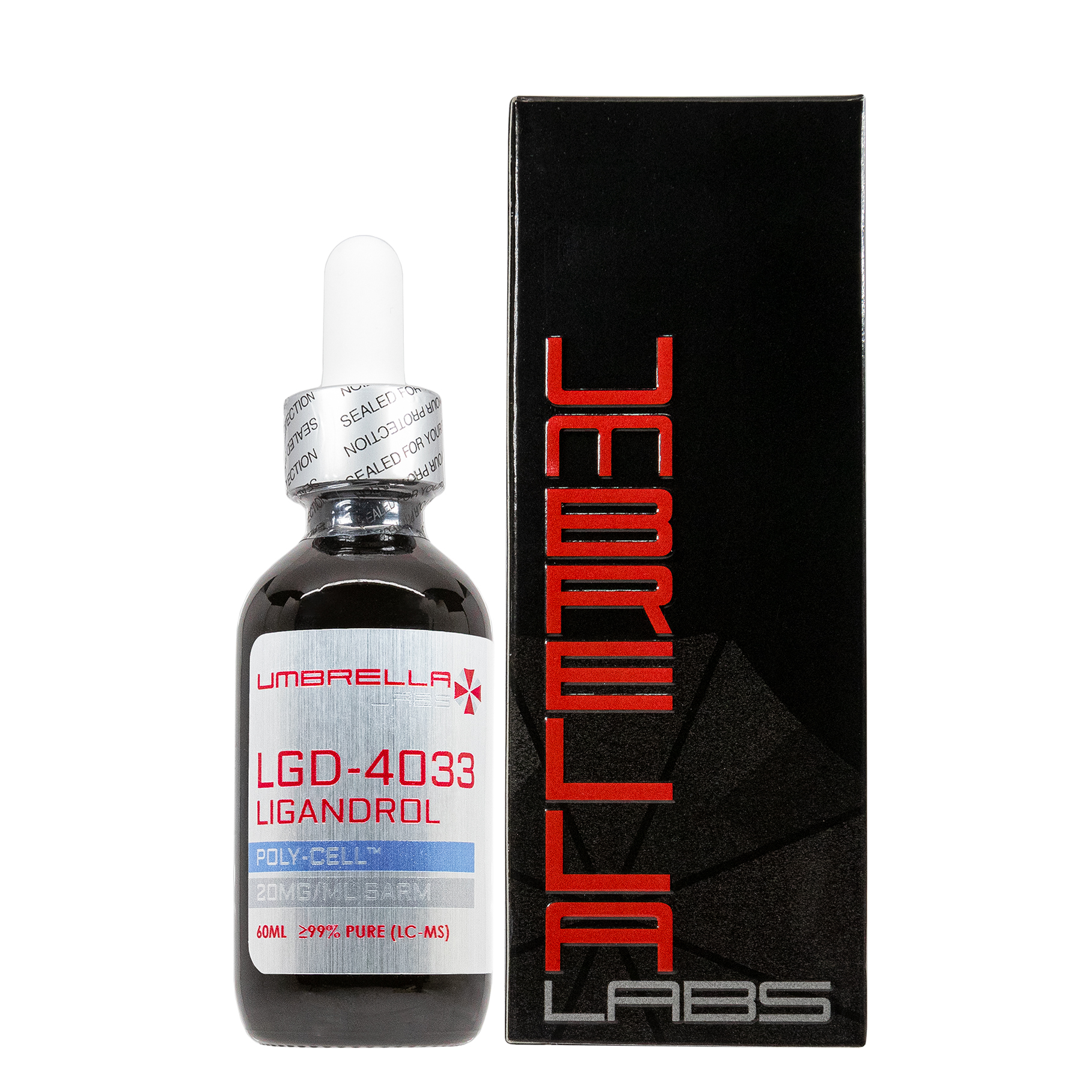
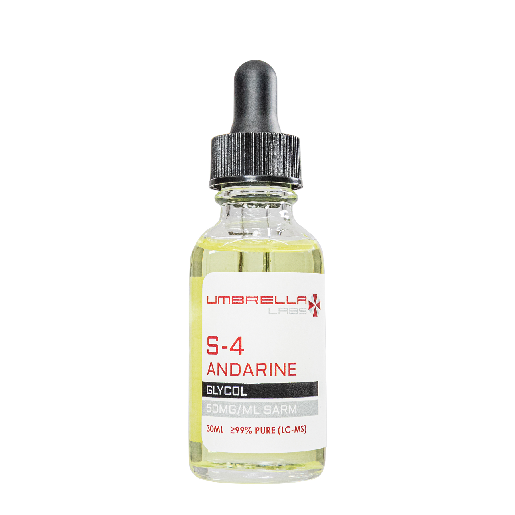
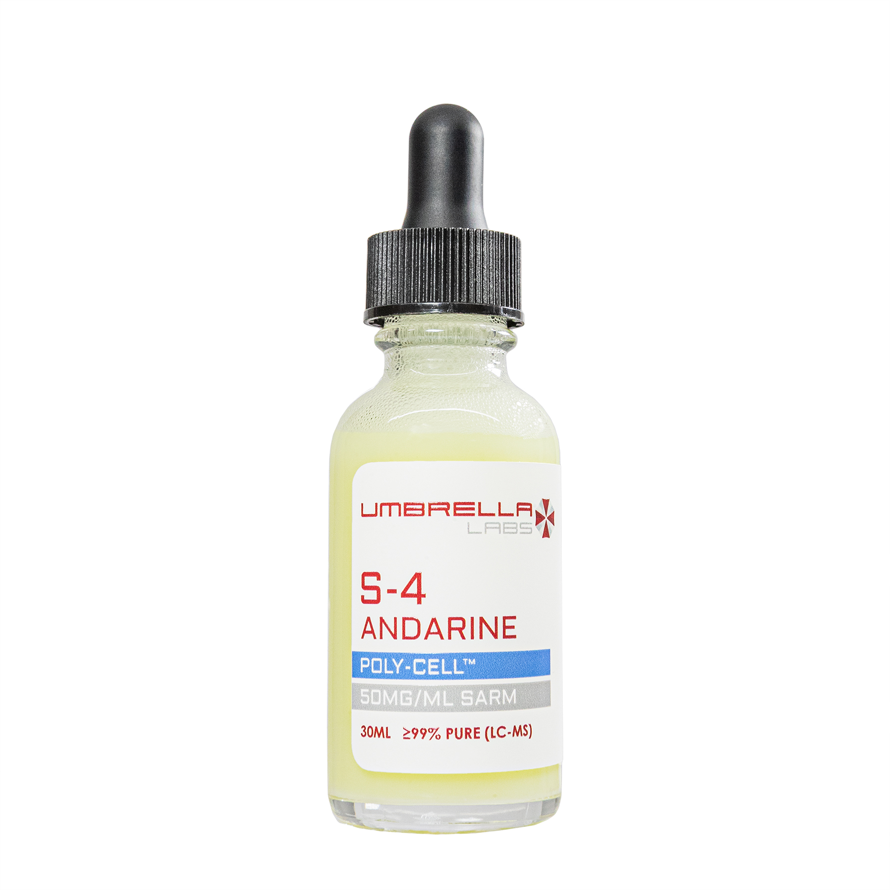
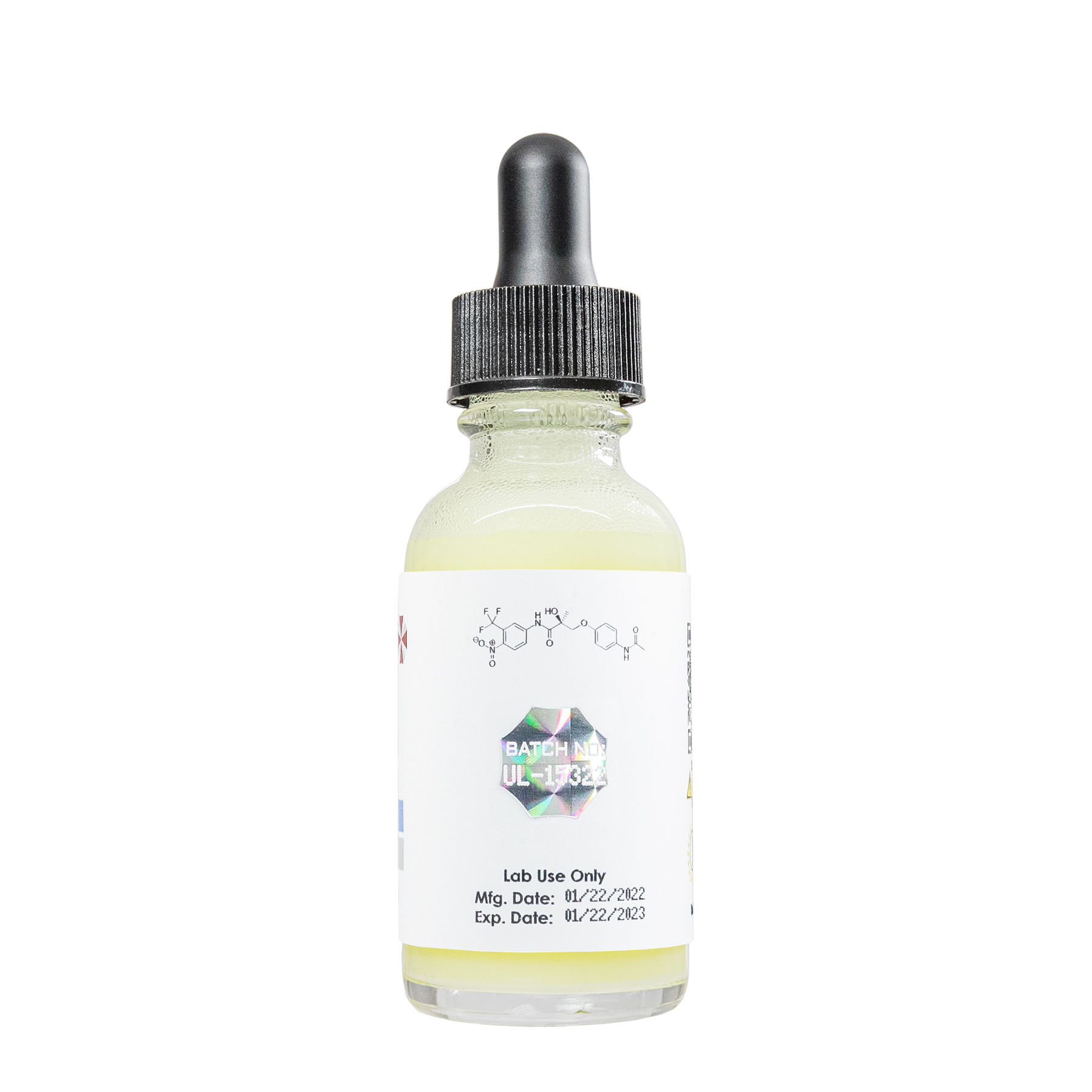
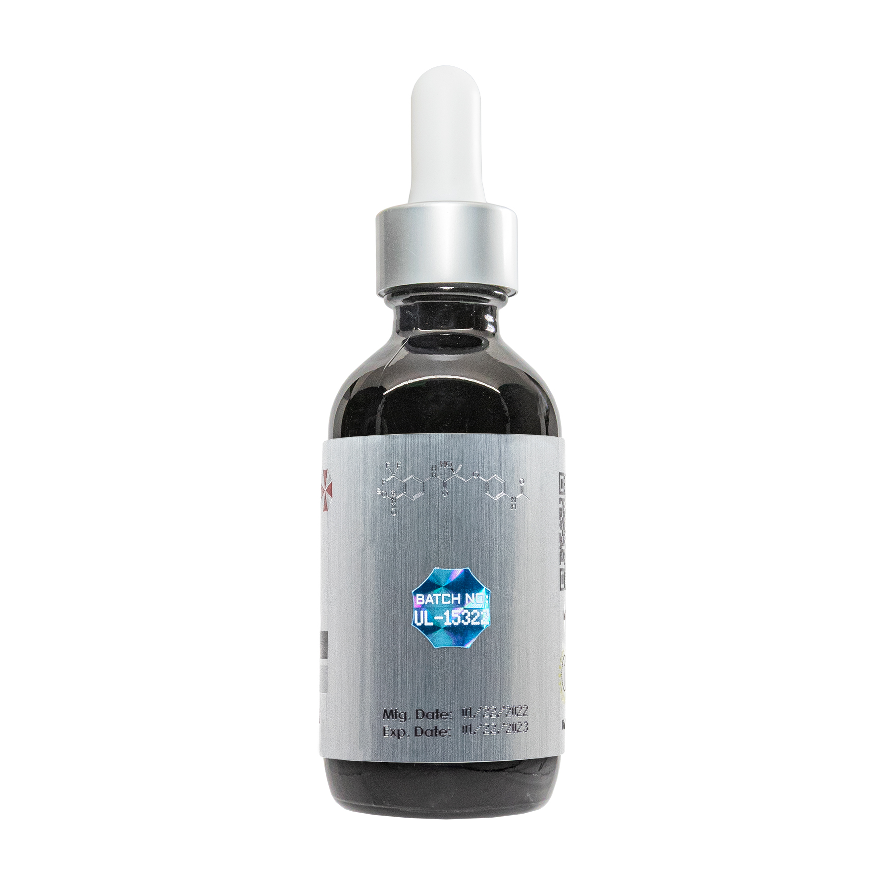
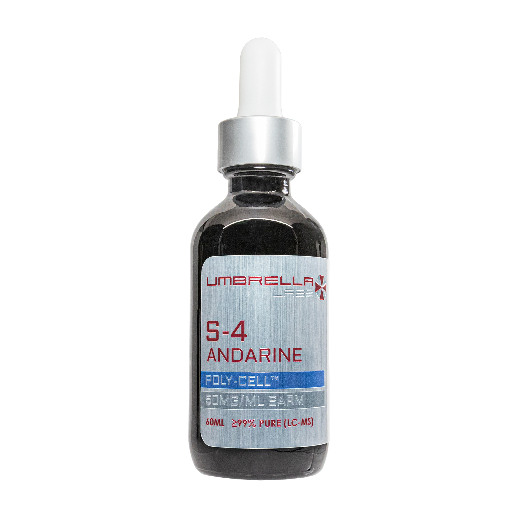
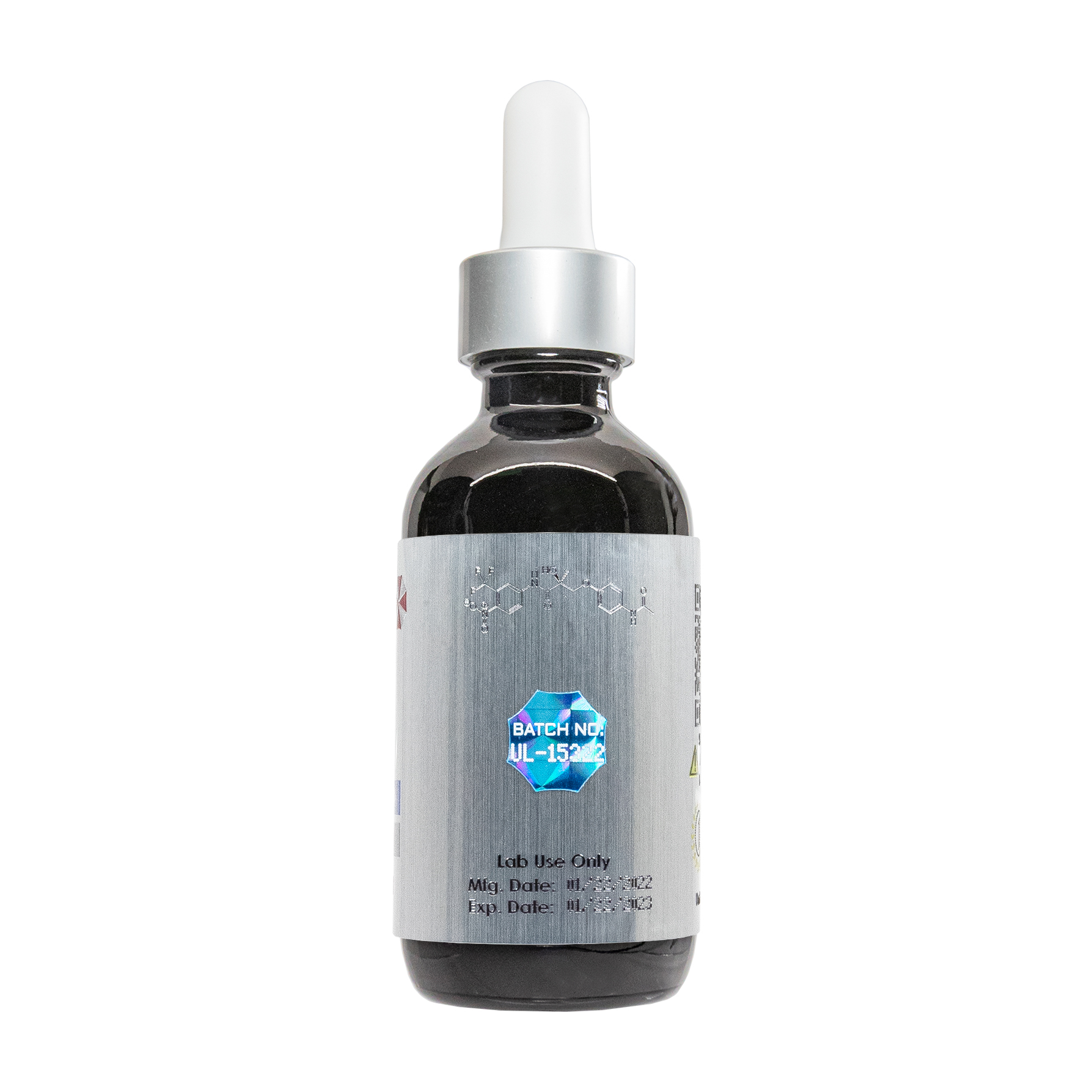
S-4 Andarine SARM Liquid
![]()
![]()
![]()
![]()
![]()
![]()
![]()
![]()

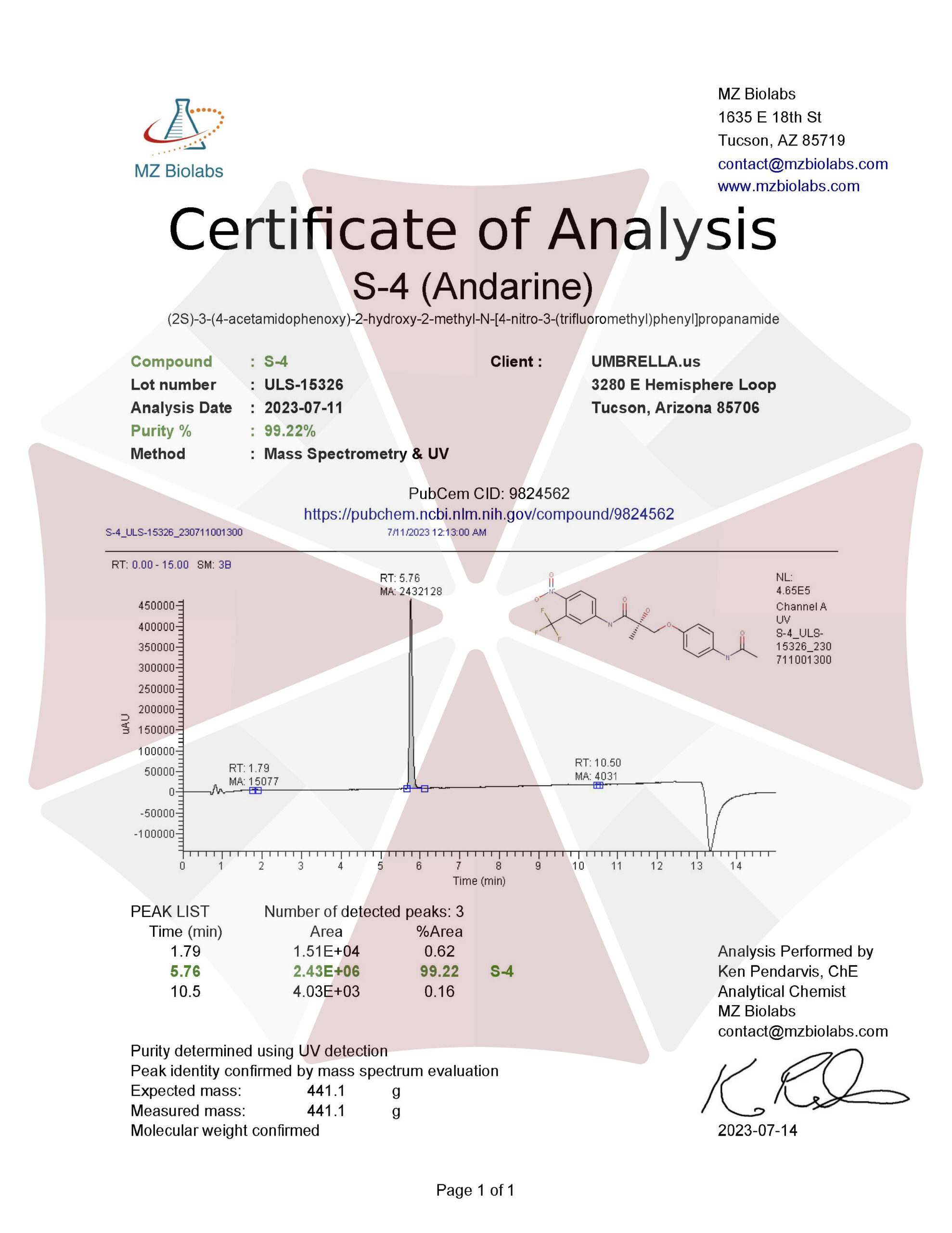
| CAS Number | 401900-40-1 |
| Other Names | S4, S-4, S 4, Andarine, GTX-007, S-4 cpd, UNII-7UT2HAH49H, 7UT2HAH49H, GTX007, GTx 007, CHEMBL125236 |
| IUPAC Name | (2S)-3-(4-acetamidophenoxy)-2-hydroxy-2-methyl-N-[4-nitro-3-(trifluoromethyl)phenyl]propanamide |
| Molecular Formula | C₁₉H₁₈F₃N₃O₆ |
| Molecular Weight | 441.4 |
| Purity | ≥99% Pure (LC-MS) |
| Liquid Availability |
|
| Powder Availability | |
| Gel Availability | |
| Storage | Store in cool dry environment, away from direct sunlight. |
| Terms | All products are for laboratory developmental research USE ONLY. Products are not for human consumption. |
What is S-4?
The selective androgen receptor modulator (SARM), S-4, commonly referred to as Andarine, is best known for its ability to promote anabolism in the bone and muscle tissues. Like other anabolic agents, S-4 binds to androgen receptors in order to initiate tissue growth, however, SARMs have been shown to cause fewer adverse side effects than typical anabolism-promoting compounds. In addition to its ability to induce growth in muscle and bone tissue, research is currently being conducted to examine the anti-cancer effects potentially elicited by Andarine.
Main Research Findings
1) Treatment with S-4 in orchidectomized rats has the potential to improve muscle strength and body composition as well as prevent bone loss.
2) Ovariectomized female rats experienced a reduction in body fat as well as a decrease in bone loss following treatment with S-4.
Selected Data
1) 12 week old male Sprague-Dawley rats were obtained from Harlan Biosciences and maintained on a strict 12 hour light, 12 hour dark cycle with food and water provided ad libitum. At the beginning of the study the test subjects were orchidectomized (ORX); a group of subjects underwent a sham-operation in order to act as a control group. The animals were randomly distributed into groups of 7 or 8 animals and allowed to recover and remain untreated for 12 weeks post-surgery in order to ensure maximum diminishment of soleus muscle strength and mass. The test subjects were then administered a 3 or 10 mg/kg dose of S-4, 3 mg/kg of DHT, or a vehicle, via subcutaneous injection, over an 8 week experimental treatment period. Throughout the treatment the animals were analyzed by DEXA scans in order to measure changes in bone mineral content (BMC, bone mineral density (BMD), lean mass, and fat mass. In order to avoid error caused by variation between days all mice were analyzed by DEXA technology at the same time [1].
At the end of the 8 week treatment period the animals were weighed and euthanized within 8 hours of the last dose. The soleus muscle of the left hind limb was extracted and analyzed further in order to determine chances in muscle strength. After muscle strength was tested the soleus muscle was frozen and preserved for electrophoretic analysis of MHC isoform expression. The heart was also dissected at the time of euthanasia and preserved to examine the MHC isoform expression in the left ventricle. Additionally, the ventral prostate, seminal vesicles, and levator ani muscle, as well as the soleus muscle from the right leg, were all extracted and weighed. Blood samples were then collected in order to measure levels of IGF-1, osteocalcin, luteinizing hormone (LH), and follicle-stimulating hormone (FSH).
As it was previously mentioned, the soleus muscle was extracted to measure muscle strength in response to androgenic treatment. Following extraction, the muscle and tendons were mounted in an experimental chamber in order for perfusion in oxygenated Krebs-Ringer solution, 137 mm NACl, 5 nM NaHCO3, 1.9 nM KH2PO4, 2 mM CaCl2, 1 mM MgSO4, and 11 mM of glucose, to occur. After the proximal and distal ends of the tendon were fixed, the muscle was stimulated through the use of a Grass S48 stimulator through two platinum field electrodes. Output measurements were recorded using ASI dynamic muscle control [1].
Furthermore, twitch kinetics and amplitude were measured while force responses were collected through stimulation of the muscle at supramaximal voltage while stretching the muscle between stimuli. Once the optimal muscle length was determined, the research team of Gao et. Al were able to measure maximal twitch, tetanic tensions, time to peak twitch tension, and time to one half twitch relaxation. Isometric twitches were elicited at 0.1 Hz; the test subjects experienced 16 continuous twitches that were recorded while one of every three twitches was analyzed. Tetanus was evoked with 3.0-sec trains of stimuli; three tetani were obtained for each muscle and the average of the the three measurements of tetanus amplitude was calculated. These procedures were followed by weighing of the soleus muscle and estimation of the cross-sectional area of the muscle [1].
In order to examine skeletal and cardiac MHC isoforms, samples of the soleus muscle and left ventricle were collected and homogenized for 5-10 seconds in sample buffer composed of 6 M urea, 2 M thiourea, 0.075 M dithiothreitol, 0.05 M Tris base, and 3% SDS. Homogenized samples were further diluted with sample buffer before they were prepared for analysis. Samples of the soleus muscle were prepared with stacking and separating gels consisting of 4 and 7% acrylamide as well as 5 and 30% glycerol, respectively, prior to the initiation of analysis. Samples of the left ventricular were similarly prepared, with the exception of the separating gel now consisting of 6% acrylamide and 5% glycerol. After analysis the samples were analyzed through the use of a scanning densitometer, provided by Hoefer Scientific. Statistical analysis was performed by single-factor ANOVA [1].
2) In the study conducted by Kearbey et. Al, the research team obtained 120 female Sprague-Dawley rats from Harlan Biosciences. The animals were given ad libitum access to food and water and were randomly housed three animals to a cage while being kept on a strict 12 hour dark, 12 hour light cycle. At 23 weeks of age the animals underwent an ovariectomy (OVX) or a sham-operation prior to random assignment into one of 12 treatment groups. The treatment groups were defined as follows: 1) OVX + S-4 (0.1 mg/day); 2) OVX + S-4 (0.3 mg/day); 3) OVX + S-4 (0.5 mg/day); 4) OVX + S-4 (0.75 mg/day); 5) OVX + S-4 (1.0 mg/day); 6) OVX + S-4 (3.0 mg/day); 7) OVX + DHT (1 mg/day); 8) OVX + S-4 (0.5 mg/day) + bicalutamide (1.0 mg/day); 9) OVX + vehicle; 10) intact + S-4 (1 mg/day); 11) intact + DHT (1 mg/day); 12) intact + vehicle. It is important to note that the researchers administered DHT treatment to both intact and OVX animals to act as a positive control group and highlight effects of a pure steroidal androgen on skeletal muscle and bone tissue. Other negative control groups were defined as intact and OVX animals receiving treatment with a vehicle, as well as a group that co-administered the antiandrogen, bicalutamide, and S-4. The purpose of the co-administration group was to delineate the AT-mediated effects in comparison to AR-independent effects of the SARM. Daily dosing solutions were prepared by dissolving the proper treatment in DMSO and diluting in PEG 300; the doses were subcutaneously injected daily, for 120 days [2].
The animals were weight and DEXA scans were performed on day 0 and day 120 in order to measure changes in total bone mineral density (BMD), percent fat mass (FM), body weight (BW), bone mineral content (BMC), bone mineral area (BMA), and lean mass (LM). Immediately after the day 120 DEXA scans, groups 1-11 were euthanized while the lumbar vertebra, femur, and tibia were extracted. Group 12 was euthanized on day 210 and bone parameters were excised and evaluated by scanning through a 3-inch deep room temperature water bath in order to simulate soft tissue. Additionally, the proximal femur, distal femur, proximal tibia, L2-L4 vertebrae, and L5-L6 vertebra were analyzed for BMD through the use of DEXA scans.
The final assessment required the research team to extract the right femurs from Group 5: OVX + 1.0 mg/day S-4, Group 6: OVX + 3.0 mg/day S-4, Group 7: OVX + 1.0 mg/day DHT, Group 9: OVX control, Group 10: intact + 1 mg/day S-4, and Group 12: intact control, in order for pQCT analysis and biomechanical testing to occur. The femur was initially analyzed at the mid-shaft and distal regions. Scout scan views were used to determine the length of the femur while the mid-shift region was chosen at 50% of the length of the femur and the distal region was chosen at 20% of the length of the femur. Additionally, a 0.5 mm perpendicular slice of the long axis of the femur was used for analytical purposes.Total and cortical BMC, BA, and BMD, as well was cortical thickness, periosteal perimeter, and endosteal perimeter were all determined at the mid-shaft. On the other hand, total and trabecular BMC, BA, and BMD were determined at the distal region of the femur [2].
Following pQCT analysis, the whole femur was used in a three-point bending test. The femur was placed on a three-point bending fixture with the anterior side of the bone facing down into an Instron Mechanical Testing Machine. The length was set accordingly while the upper loading device was aligned to the center of the shaft in order for a load to be applied at a constant displacement until the femur broke. The testing machine measured maximum load, stiffness, and energy absorbed. The research team calculated the axial area moment of inertia from the data collected from the pQCT analysis. Finally, statistical analyses were performed by Fisher’s Protected Least Significant Difference test for multiple comparisons [2].
Discussion
1) Initial results of the study conducted by Gao et. Al reported that 20 weeks after the rats underwent orchidectomy, the body weight and soleus muscle had significantly decreased in comparison to the intact control animals. Androgenic treatment with either S-4 or DHT led to a slight increase in body and muscle weight in the ORX animals, however, the increase was deemed insignificant. Following ORX, the length at which maximal twitch tension was elicited (L0) was significantly reduced as well, however, S-4 and DHT returned L0 back to the same level as the intact animals. There was no noticeable difference in CSA between the treatment groups. Due to the findings of this study, the research team was able to conclude that diminishment in soleus muscle weight and L0 was likely due to the decrease in animal body size. Additionally, because S-4 and DHT treatment did not significantly increase soleus muscle weight, previous findings regarding the lack of androgen-sensitivity of the soleus were confirmed. This was compared to the levator ani muscle that has been shown to be highly androgen-sensitive; weight of the levator ani decreased by 64% while soleus weight only decreased by 11% after ORX [1].
Peak tetanic tension was measured as the contractile force of the soleus muscle. Androgen treatment with S-4 and DHT did not increase the muscle size but rather led to a significant increase in the peak tetanic tension of the soleus muscle in ORX animals. Following ORX, peak tetanic tension of the soleus decreased from 0.85 n to 0.57 n. When 3 mg/kg of S-4 and 3 mg/kg of DHT were administered, peak tetanic tension increased to 0.86 n and 0.95 n, respectively. 10 mg/kg of S-4 increased peak tetanic tension to 1.02 n, which was higher than the intact animals.
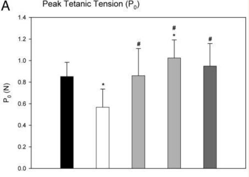
Figure 1: Changes in peak tetanic tension in response to various treatments
Tetanus and twitch parameters did not differ significantly among ORX, S-4, and DHT treatment groups. Only higher doses (10 mg/kg and up) of S-4 increases peak twitch tension in ORX animals. Overall, the initial results found that ORX decreased the body weight, soleus muscle weight, L0 and peak tetanic tension in rats. S-4 and DHT increased strength of the soleus muscle (P0) of ORX animals compared to intact animals. While androgen treatments also showed potential in increasing soleus muscle mass in ORX animals, however, the change was not significant [1].
The next portion of the study examined how S-4 and DHT restored androgen-dependent tissues followed by androgen deprivation induced by orchidectomy. The animals were left untreated to ensure proper atrophy which resulted in the prostate seminal vesicles, and levator ani muscle decreasing to 3.6, 6.7, and 41.4%, respectively, of the intact animals. The weight of the prostate and seminal vesicles doubled after treatment with DHT while S-4 restored the weight of the levator ani but not the prostate and seminal vesicles to less than 20% of the intact control groups. Increasing doses of S-4 continued to increase weight of the levator ani and elicited stronger androgenic effects in the prostate and seminal vesicles. This demonstrated that S-4 can restore androgen-dependent tissue after androgen depletion [1].
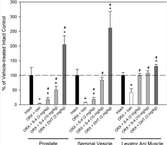
Figure 2: weight of the prostate seminal vesicles, and levator ani in response to different treatments
The research team measured plasma levels of IGF-1 and osteocalcin as these compounds are typically used as markers of anabolic activity and bone turnover rate. IGF-1 levels in ORX animals 20 weeks after surgery did not significantly differ from IGF-1 levels in intact animals. S-4 did not lead to any dramatic changes in IGF-1 levels while DHT decreased IGF-1 concentration to approximately 70% of the level observed in intact animals. In terms of osteocalcin, plasma levels were not drastically different between intact and ORX test subjects, 20 weeks after orchiectomy. 8 weeks of treatment with 3 or 10 mg/kg of S-4 or 3 mg/kg of DHT in ORX levels led to a decrease in plasma osteocalcin levels in 70% and 50%, respectively, of the intact animals. This suggests decreased bone breakdown in ORX animals in response to androgenic treatment [1].
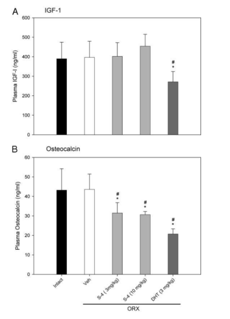
Figure 3: Plasma IGF-1 and osteocalcin levels in response to different treatments
DEXA scans were used to record the effects of DHT and S-4 on muscle and body tissues. BMD and BMC were significantly lower than the intact animals following orchidectomy. The animals were then treated with a vehicle, 3 or 10 mg/kg of S-4, or 3 mg/kg of DHT for 8 weeks. BMD and BMC in vehicle-treated intact animals increase by 1.45 and 12.92 grams, respectively, while ORX animals experienced increases in BMD and BMC by 0.65 and 11.18 grams, respectively. There were no dramatic differences in weight among the ORX groups, however, S-4-treated ORX animals experienced an increase in BMD compared to vehicle-treated ORX animals. The increase in BMD was similar to the intact animals. The 10 mg/kg dose of S-4 also led to a significant increase in BMC in a manner similar to intact animals, measuring at 12.00 grams. DHT led to increased BMD and BMC, however, the test subjects did not exhibit changes as significant as the group treated with both doses of S-4. Additionally, the test subjects did not experience any significant weight changes, but out of all groups the S-4 treated ORX animals gained more weight than the ORX animals treated with a vehicle. S-4 did not decrease fat mass either, but treatment did restore body composition to a manner similar to intact animals [1].
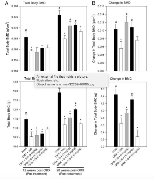
Figure 4: changes in total body BMD and BMC in response to different treatments.
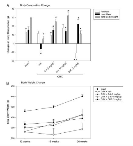
Figure 5: changes in body composition in response to different treatments
Finally, the researchers attempted to explain why muscle strength increased but muscle size did not in response to androgen treatment. SDS-PAGE analysis was used to examine the expression of MHC isoforms in soleus and cardiac muscle samples. In the soleus muscle sample, two isoforms were detected: MHC-I and MHC-IIa. MHC-I accounted for more than 85% of the total MHC expressed. Seven out of eight samples in ORX animals expressed MHC-IIa where only two of the seven samples in intact animals expressed MHC-IIa. Because there was no significant differences between the treatment groups, the slow-to-fast shift observed in ORX animals did not account for muscle strength
In terms of cardiac muscle, the two main MHC isoforms expressed with MHC-alpha and MHC-beta. MHC-alpha accounted for 65% of total MHC expressed in intact animals, however MHC-alpha expression decreased to 44% in ORX animals. 3 mg/kg of S-4 restored MHC-alpha expression to 57% in ORX animals. While both S-4 and DHT restored MHC-alpha expression significantly, expression did not reach the same levels as intact animals. The beta-to-alpha shift in cardiac muscle is regulated by androgens and is related to androgen function in the heart. However, the increase in muscle strength induced by androgen treatment was not related to the slow-to-fast shift in the soleus muscle [1[.
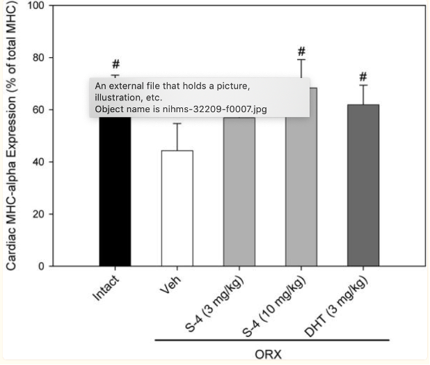
Figure 6: Expression of cardiac MHC alpha and beta isoforms in response to different treatment
2) Results of the study conducted by the research team of Kearby et. Al determined that ovariectomy leads to decreased BMD in comparison to the intact control group. Treatment with 0.1 mg/kg doses of S-4 led to partial or full prevention of BMD loss in OVX animals. When S-4 was administered with the anti-androgen, bicalutamide, the effects elicited by the SARM were prevented. This indicated that the androgen receptor is crucial to regulation of bone response to the drug. Additionally, body weight and body composition was observed through DEXA scans. OVX groups exhibited greater body weight than the intact groups, suggestions that animal growth is influenced by estrogen-deprivation. Treatment with 3 mg/day of S-4 led to further increases in body weight in OVX animals, however S-4 administration in intact animals resulted in a decrease in body weight in comparison to OVX and intact controls.
Percent fat mass was also measured through DEXA scans. The OVX control exhibited higher levels of FM than intact control groups. Additionally, S-4 treatment resulted in dose-dependent decrease in FM to a similar level to the intact controls. Vertebral bone loss was also measured by DEXA analysis. L5YL6 vertebrae were extracted to observe vertebral BMD. S-4 treatment led to a dose-dependent reduction in bone loss. The 3 mg/day dose prevented bone loss completely while the 0.5 and 1 mg/day doses partially prevented bone loss. The elicited effects were inhibited when the anti-androgen bicalutamide was administered in conjunction with S-4 [2].
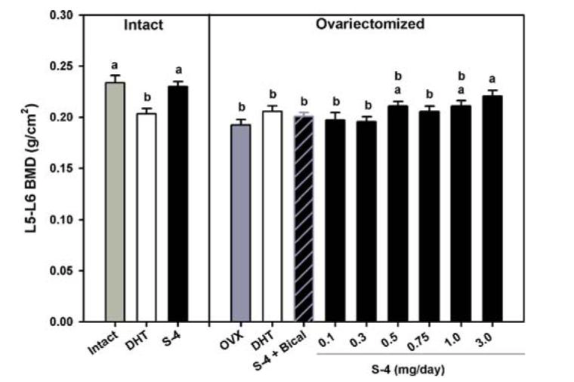
Figure 7: Changes in BMD in response to different treatments
The femurs of the rats were analyzed by pQCT at the mid-shaft for cortical thickness, periosteal circumference, cortical content, and cortical density, and at the distal femur for trabecular BMD. Following OVX, S-4 treatment led to an increase in cortical thickness above the levels reported in the intact control groups. Additionally, S-4 was able to completely inhibit the loss of cortical content at the femoral midshaft after OVX was completed. The 3 mg/day dose of S-4 led to an increase in cortical content, however, the change was deemed insignificant, however, S-4 treatment in OVX rats prevented a decrease in periosteal circumference. While cortical thickness was increased in intact animals administered S-4, periosteal circumference was decreased.
pQCt was also used to measure cortical bone mineral density (CD) of the femoral mid-shaft; S-4 treatment prevented the OVX-induced loss of CD. In comparison to OVX and intact controls, CD was increased in intact animals receiving 1 mg/day of S-4. OVX also led to significant trabecular bone loss in the distal femur. S-4 partially prevented this loss; in intact animals S-4 treatment was shown to increase trabecular BMD to a higher level than the intact controls. This indicates that S-4 promotes anabolic activity in the trabecular bone regions. Furthermore, a three-point bending analysis was used to measure biomechanics strength. S-4 treatment in OVX animals was found to prevent the loss of femoral biomechanical strength [2].
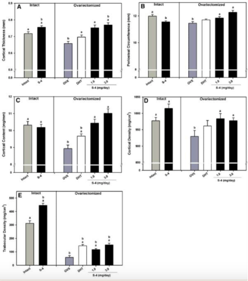
Figure 8: QCT analysis of the femur. A) cortical thickness, B) periosteal circumference, C) cortical content, D) cortical bone density, E) trabecular density
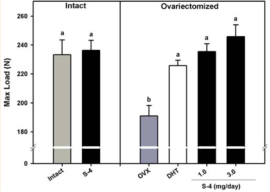
Figure 9: Maximum load determined by three-point bending evaluation.
Disclaimer
**LAB USE ONLY**
*This information is for educational purposes only and does not constitute medical advice. THE PRODUCTS DESCRIBED HEREIN ARE FOR RESEARCH USE ONLY. All clinical research must be conducted with oversight from the appropriate Institutional Review Board (IRB). All preclinical research must be conducted with oversight from the appropriate Institutional Animal Care and Use Committee (IACUC) following the guidelines of the Animal Welfare Act (AWA).
Citations
[1] Gao W, Reiser PJ, Coss CC, Phelps MA, Kearbey JD, Miller DD, Dalton JT. Selective androgen receptor modulator treatment improves muscle strength and body composition and prevents bone loss in orchidectomized rats. Endocrinology. 2005 Nov;146(11):4887-97. doi: 10.1210/en.2005-0572. Epub 2005 Aug 11. PMID: 16099859; PMCID: PMC2039881.
[2] Kearbey JD, Gao W, Narayanan R, Fisher SJ, Wu D, Miller DD, Dalton JT. Selective Androgen Receptor Modulator (SARM) treatment prevents bone loss and reduces body fat in ovariectomized rats. Pharm Res. 2007 Feb;24(2):328-35. doi: 10.1007/s11095-006-9152-9. Epub 2006 Oct 25. PMID: 17063395; PMCID: PMC2039878.
S-4 Andarine SARM is sold for laboratory research use only. Terms of sale apply. Not for human consumption, nor medical, veterinary, or household uses. Please familiarize yourself with our Terms & Conditions prior to ordering.


| File Name | View/Download |
| 07-11-2023-Umbrella-Labs-S-4-Andarine-Certificate-Of-Analysis-COA.pdf |
VIEW CERTIFICATES OF ANALYSIS (COA)
Additional information
| Weight | 4 oz |
|---|---|
| Dimensions | 4 × 4 × 2 in |
| Formula Option | |
| CAS Number | |
| PubChem CID | |
| ChEMBL ID | |
| ChemSpider ID | |
| Molar Mass | 441.363 g·mol−1 |
| Molecular Formula | C19H18F3N3O6 |
| Size |